An open-source textbook for college-level students and educators
11 Non-Opaque Oxides
11.1 Rutile
TiO2
Occurrence—Rutile is a common accessory mineral in metamorphic schists and gneisses. It also is found as small grains in intermediate to mafic igneous rocks. Less common occurrences are in veins, in pegmatites, and in some sediments.
Distinguishing Features—Rutile typically occurs as small grains, or prismatic or needle-like crystals. Simple twins forming “kinks” are common in pegmatites but are rare in other rocks. A strong red, reddish-brown, or amber color is a key to identification (see, for example Figs 11.1.1 and 11.1.2), but rutile may also be yellowish or yellow-brown or less commonly purple-blue. Fine needles may be pale.
Crystals have very-high relief. They show extreme birefringence that may be difficult to see because of the strong color of the crystals. Rutile has good cleavages that are rarely visible in thin section except in end sections.
Similar Minerals—Rutile’s red color may sometimes resemble the color of (thin) hematite or iron oxyhydroxides like goethite or lepidocrocite. It may also be confused with cassiterite (lower relief and birefringence), titanite (colorless), or brookite.
rutile
Optical Properties ■■ Tetragonal; uniaxial (+) ■■ ω = 2.609-2.616, ε = 2.895-2.903 ■■ δ = 0.286-0.287. Interference figures show many isochromes ■■ Rutile has parallel extinction ■■ Views down the c-axis show cleavages intersecting at 45o
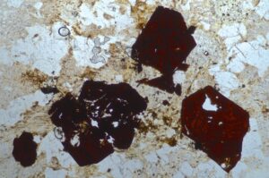
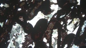
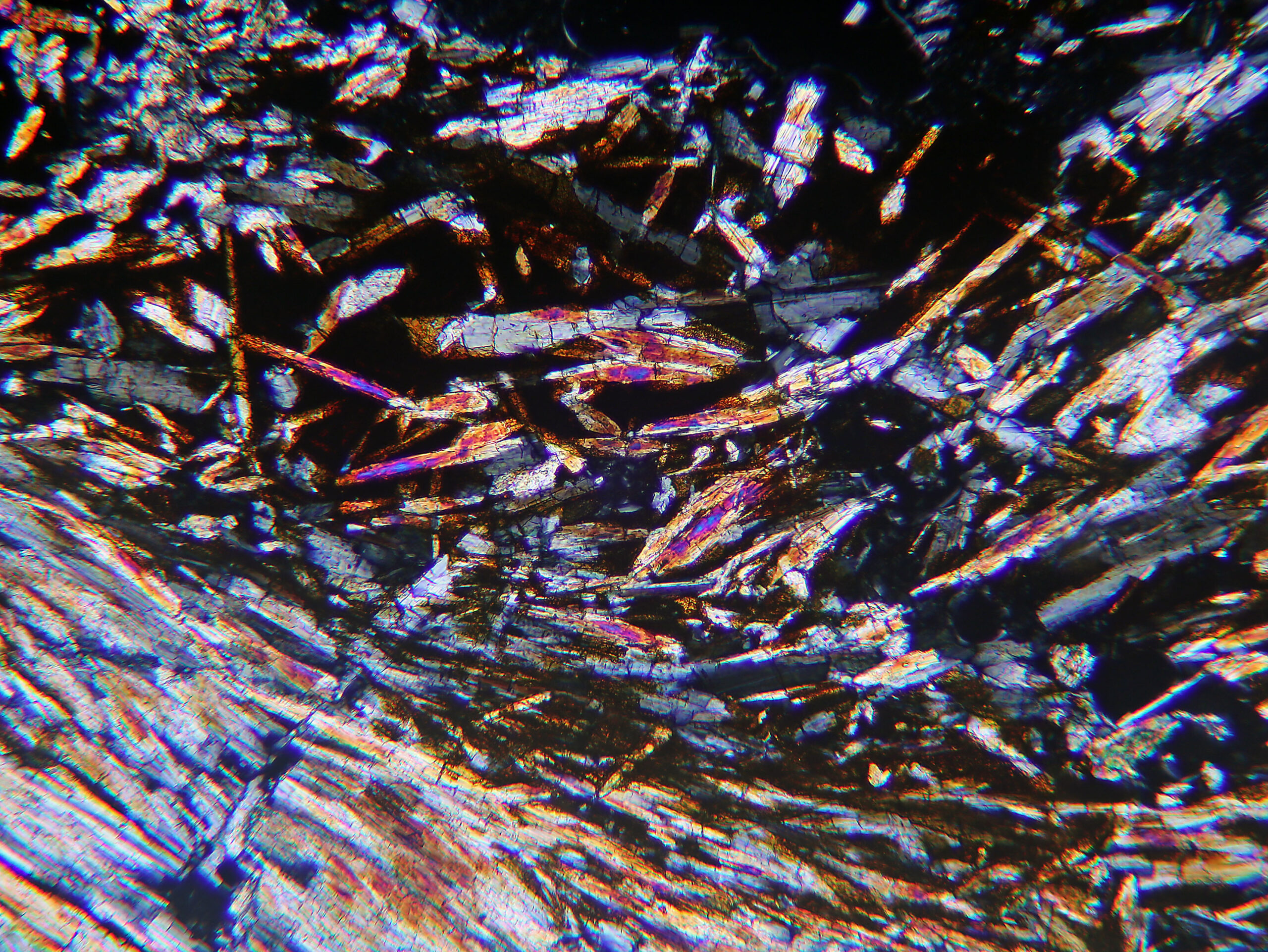
Fig 11.1.1 Rutile
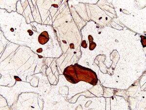
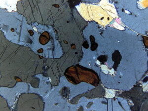
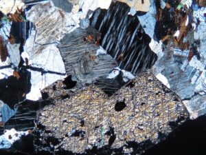
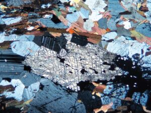
This thin section contains very high-relief reddish-brown rutile. The rutile is accompanied by light-tan gedrite (which shows classic amphibole cleavage) and lower-relief cordierite. In the XP view, rutile’s interference colors cannot be seen due to the deep color of the mineral. Sometimes, however, rutile interference colors can be seen on very thin grain edges. End views of the gedrite show very low-order interference colors because the view is close to an optic axis; the one gedrite grain in longitudinal section shows 1st-order yellow colors. The cordierite shows its typical 1st-order gray colors. It does not display the twinning or pleochroic halos that are often keys to cordierite identification. Photos from Dr. Kurt Hollocher. FOV = 1.2 mm.
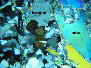
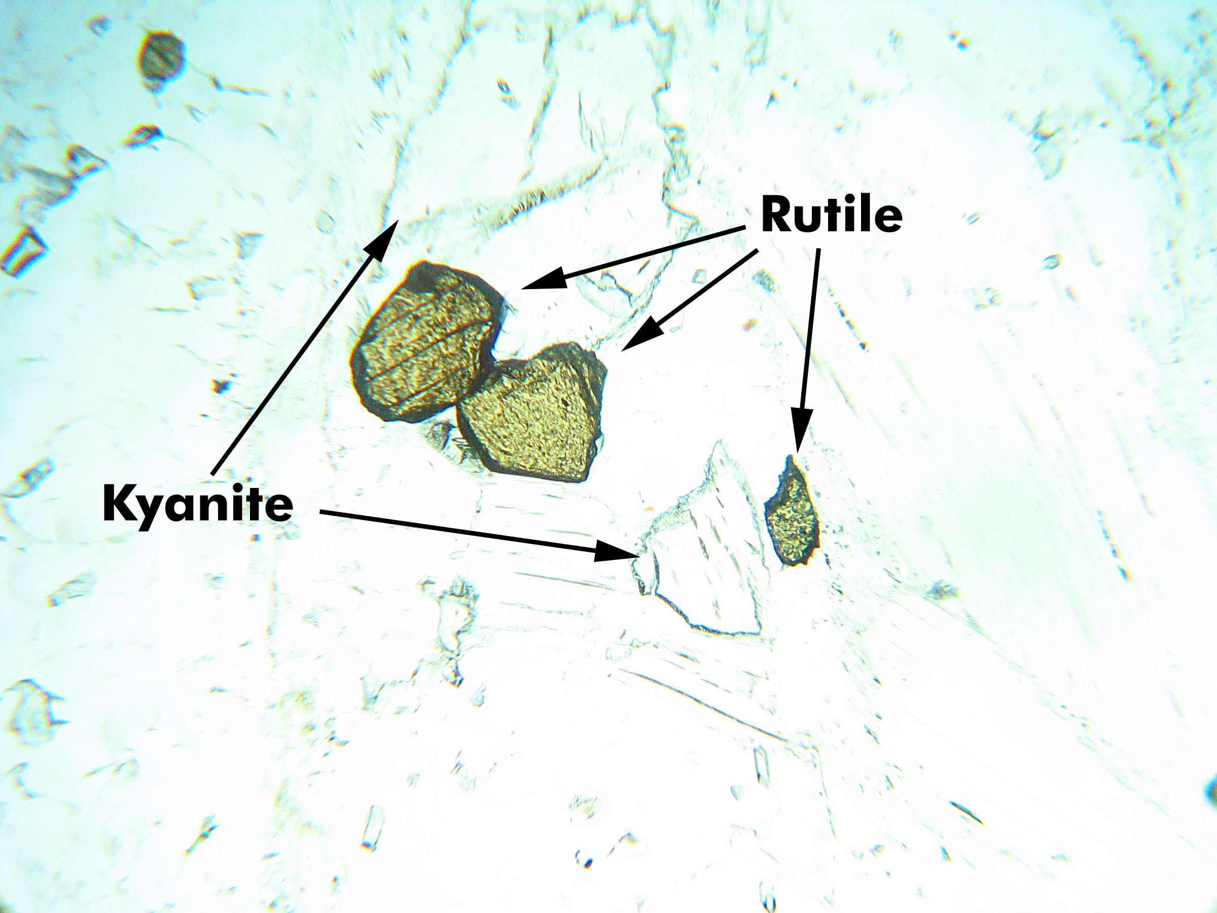
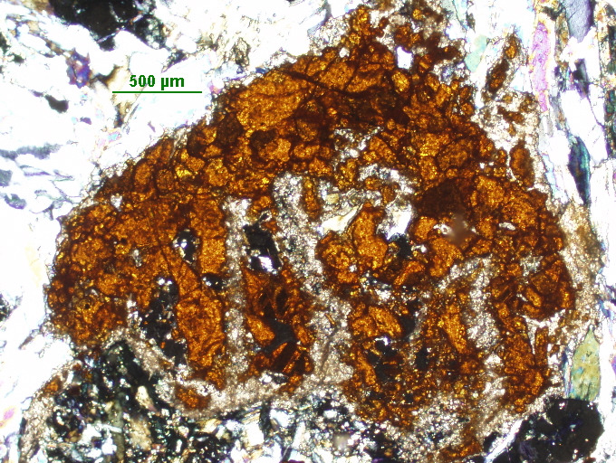
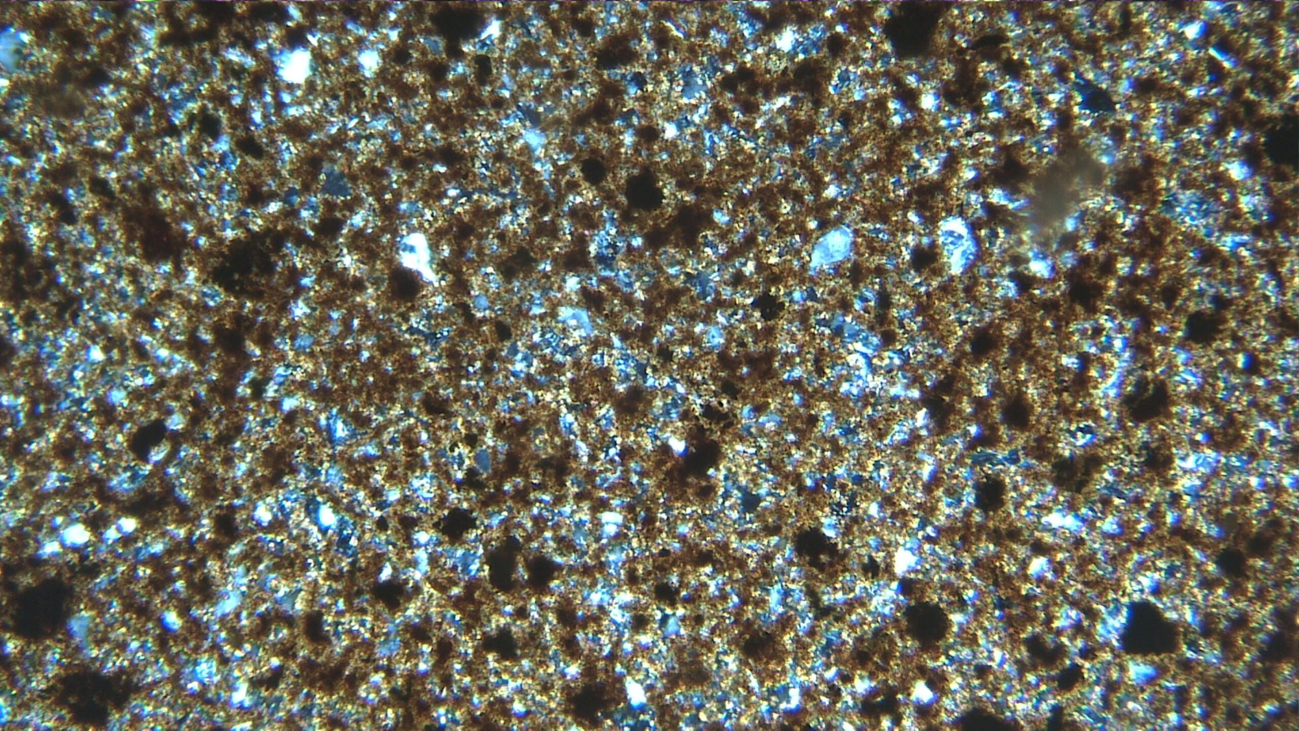
This view shows several yellow-tan rutile grains center of view) in a whiteschist from the Dora Maira Massif, Italy. The rutile is surrounded by mostly quartz (on the left and bottom of the photo) and several flakes of clear mica (on the right). Rutile has very high birefringence. Just a hint of high-order pastel interference colors can be seen here (XP). Although not distinctive in these photos, kyanite is present just above the two large rutile grains, and also to the left of the smaller grain near the center of the field of view. The kyanite has slightly higher relief than surrounding minerals, but otherwise is hard to pick out. It is clear (PP) and shows 1st-order gray interference colors (XP). This rock also contains lots of quartz. The field of view is 1.5 mm across.
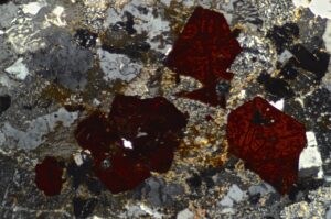
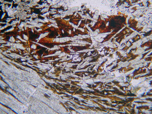
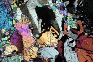
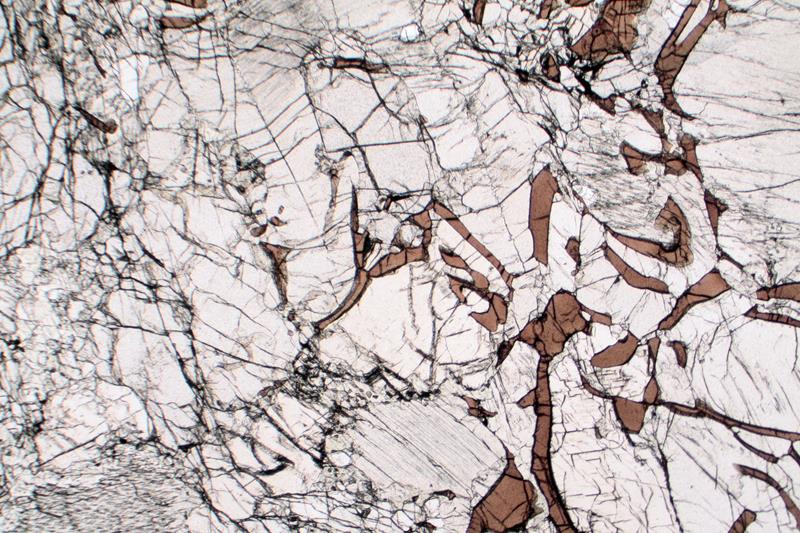
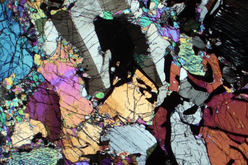
This schist contains many grains of high-relief brown-red rutile in a sea of mostly muscovite. Opaque-appearing rims are fine-grained titanite. The relatively clear, high-relief, tabular grains are zoisite. In the XP view, rutile’s high-order interference color almost show in a few places but are mostly masked by the mineral color. Muscovite displays typical 2nd-order colors, and zoisite displays anomalous royal blue interference colors. Photos from science.smith.edu. FOV = 2.5 mm.
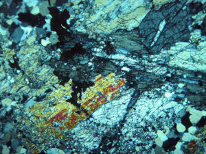
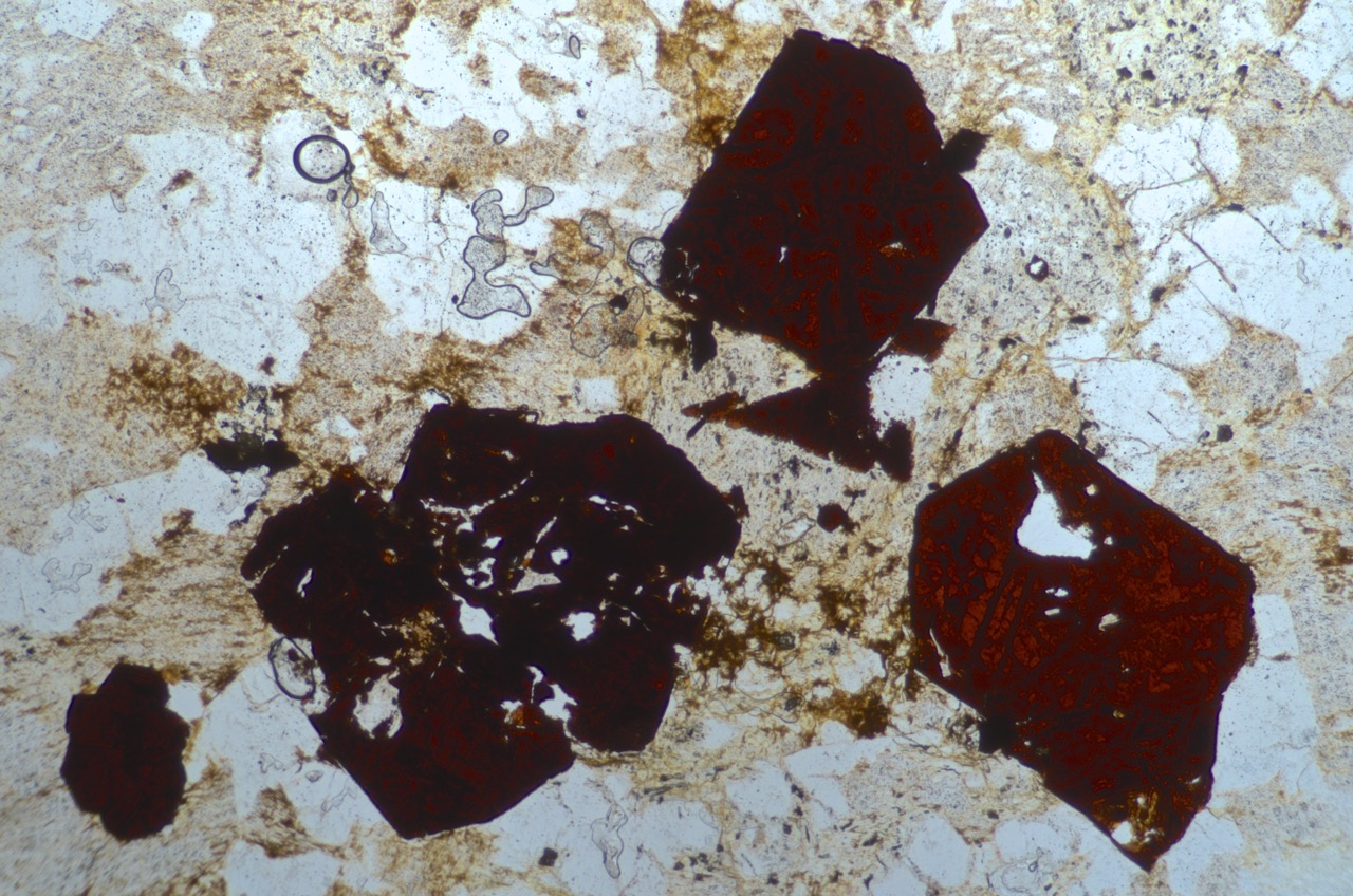
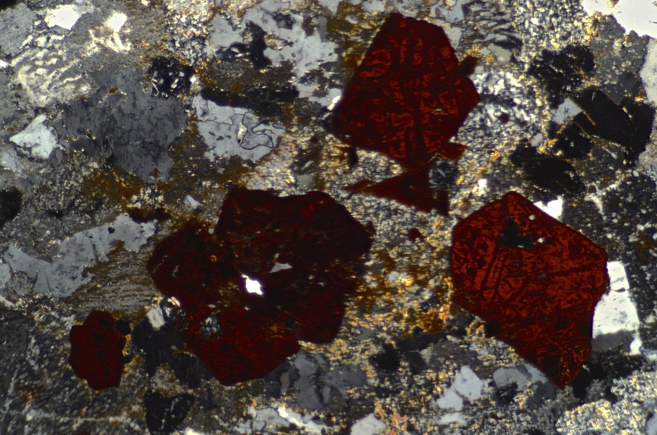
The dark mineral, not quite isotropic in this view, is rutile. At higher magnification with conoscopic light, its reddish-brown color would be evident. Most of the section, however, is kyanite (high relief, cracked/cleaved/fractured). The kyanite shows good cleavage, and some grains are twinned. In the XP view, rutile shows no interference colors; the kyanite displays up to 2nd-order yellow. Minor quartz is present, most notably in the lower right corner of the view. This view is about 1 mm across.
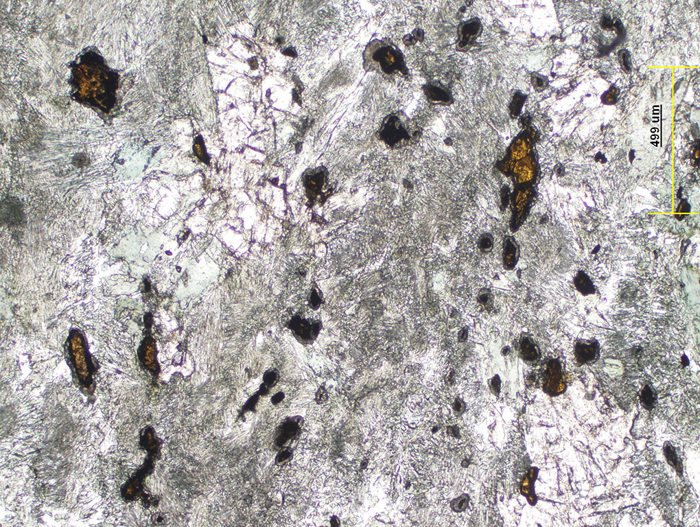
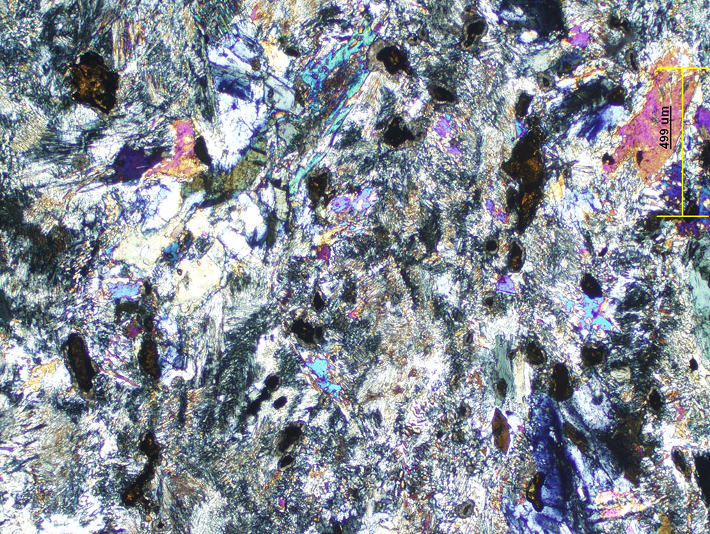
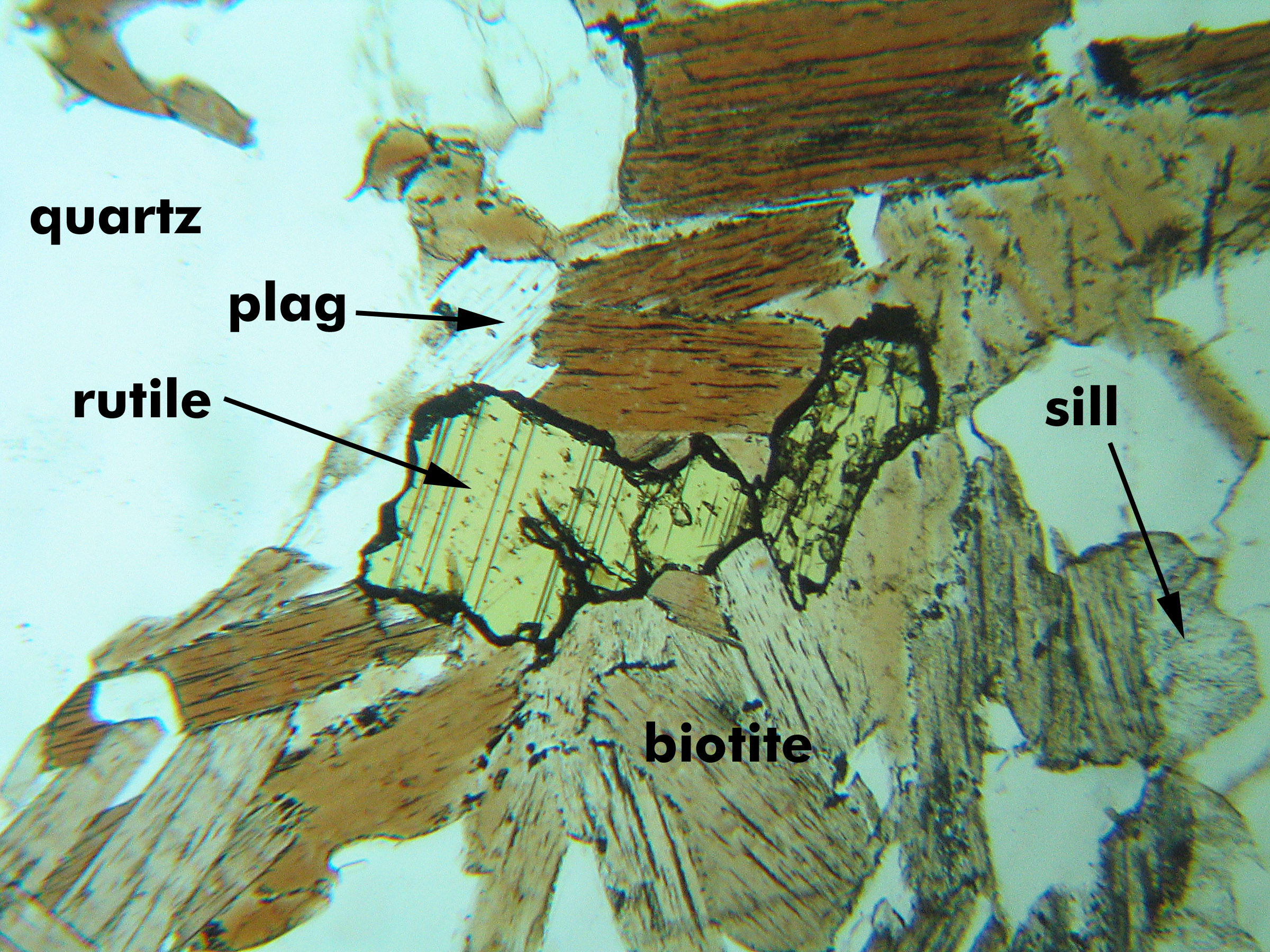
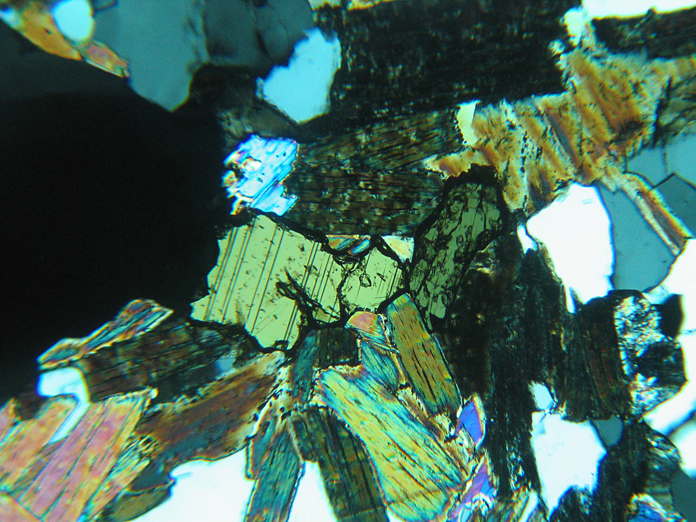
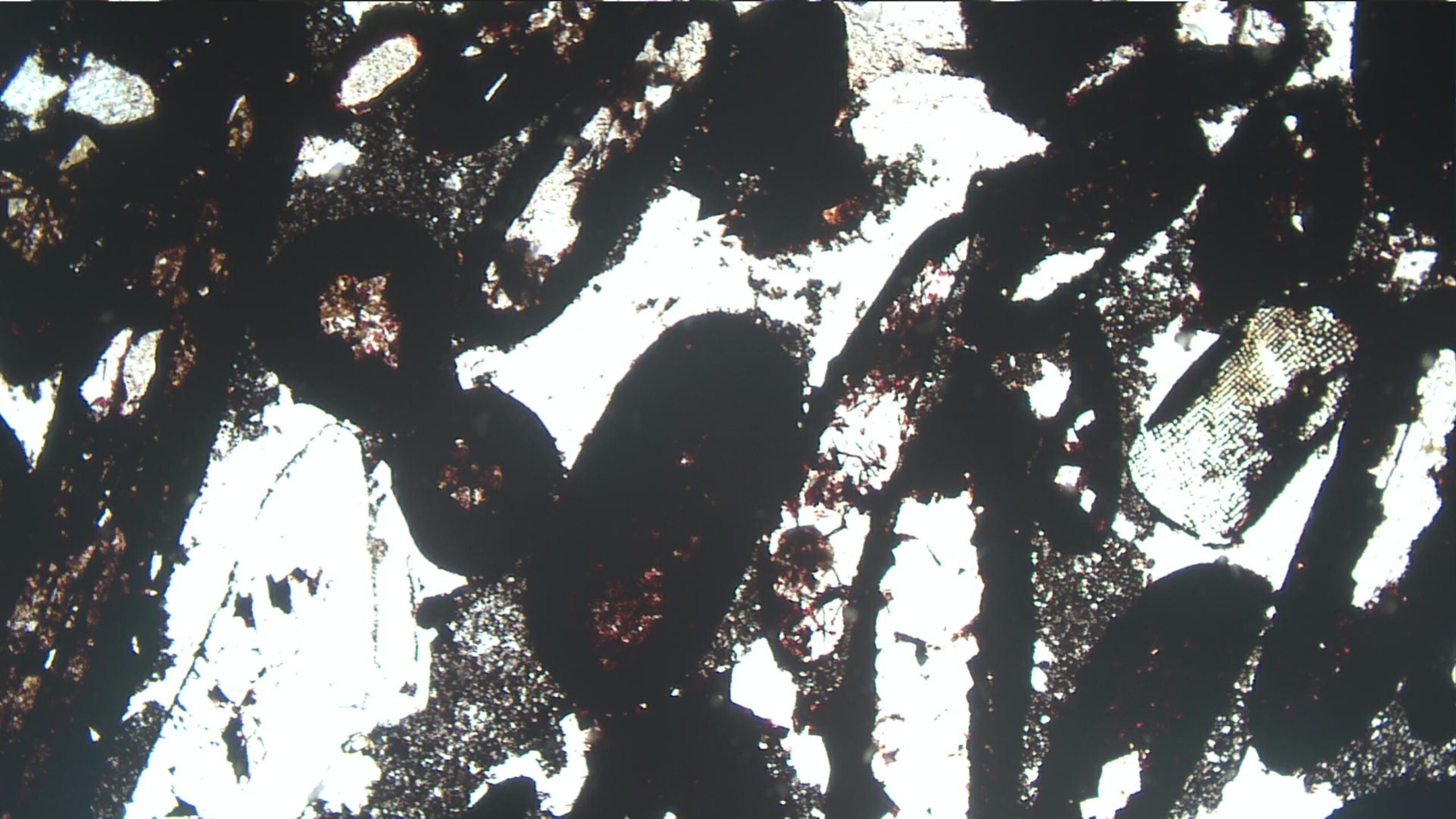
This is a biotite-sillimanite-rutile schist from Montana’s Beartooth Mountains. The yellowish rutile is twinned, and the twins show in both the PP and XP views. The rutile is rimmed by an opaque mineral, likely ilmenite. The biotite is flaky and pleochroic in various shades of brown; it shows up to high 2nd-order interference colors. Quartz is also a present (clear grains with low birefringence), and one grain of plagioclase. FOV = 1.5 mm.
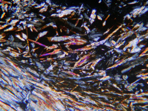
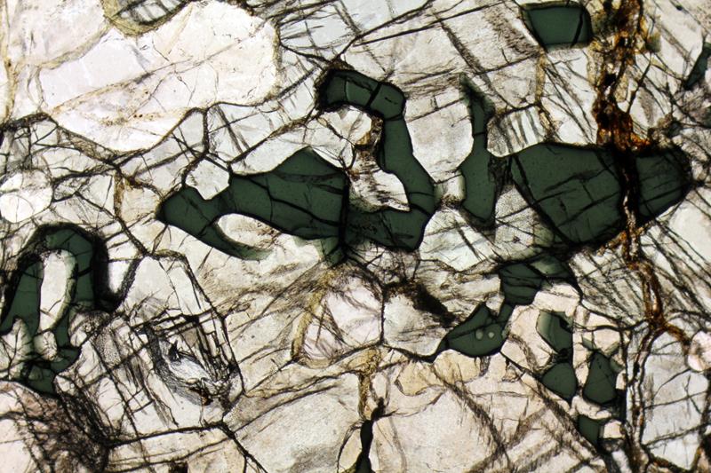
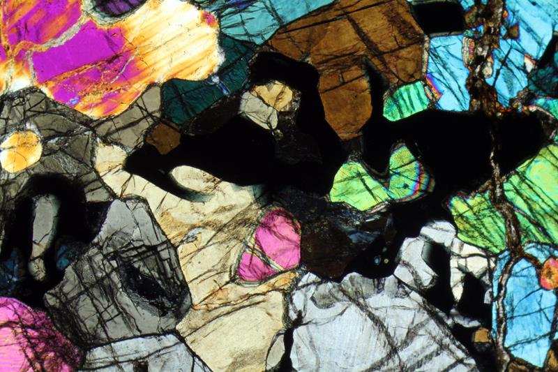
This view shows a mass of anhedral reddish rutile crystals in a blueschist from Syros, Greece. Although rutile has very high birefringence, rutile’s color appears the same in both the PP and the XP view because of the strong color of the mineral. The high-relief colorless to tan mineral on edges of rutile is titanite. Photos from science.smith.edu. FOV = 3.5 mm.
Occurrence—Hematite is a common mineral in diverse rocks, notably as a cement in red sandstone. It can form as an alteration product after just about any Fe-bearing mineral.
Distinguishing Features—Red or brown-red color is a key identifier. Hematite crystals display high relief and may be euhedral or form masses or clots of fine-grained aggregates. They do not display cleavage but commonly fracture.
Coarse crystals are normally opaque, but thin crystals may show some red or yellow to brown-red color, especially at their edges (most noticeable with the conoscopic lens inserted). Twinning is common but rarely seen in thin section.
Interference colors are very high but masked by the color of the crystals.
Similar Minerals—Hematite is similar to goethite and lepidocrocite, common secondary iron hydroxides, but goethite and lepidocrocite are lighter colored and more brownish. Ilmenite is darker and blacker.
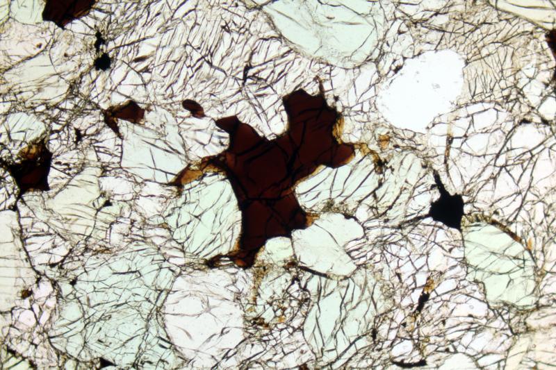
This is a view of one member (hematite sandstone) of the Clinton Iron Formation from northwestern New York. The brown material is mostly hematite, some hydrated to limonite, but distinguishing hematite from limonite is problematic in this thin section view. Clear grains are quartz (PP). In the XP view, the dark color of the hematite persists, and the quartz shows typical 1st-order colors. FOV = 1.5 mm.
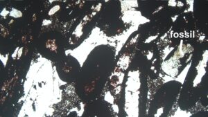
This rock contains dark-brown deformed hematite oolites, pellets with shale cores and hematite rims, clear calcite, and at least one fossil. In the XP view, the hematitic grains remain brown and the calcite shows very-high order pearl-white interference colors. This rock is also from the Clinton Iron Formation, like the previous one, but is a limestone instead of a sandstone. FOV = 3.5 mm.
This is taconite, a type or low-grade iron ore, from Hoyt Lakes, Minnesota. The rock contains clear quartz grains with dark-brown hematite rims (PP). The hematite is probably mixed with limonite or some other Fe-hydroxide. The mostly clear interstitial material is calcite. In the XP view, the hematite appears dark brown, quartz shows its usual interference colors, and the calcite displays high-order and pearl-white colors. FOV = 3.5 mm.
The clear to very pale green mineral in this image is actinolite. Orangish iron oxide occurs between the amphibole crystals, likely replacing calcite. Hematite can be translucent like the orange oxide in this image, but the orange material may contain other iron oxides or hydroxide, too. This rock comes from the Ruby Mountains, Montana. FOV = 1.25 mm.
Occurrence—Corundum can be found as an accessory mineral in metamorphosed carbonates, in highly aluminous metasediments, in some Al-rich igneous rocks, and in placers. Massive corundum is found in skarns.
Distinguishing Features—Corundum is generally colorless in thin section and has very high relief. Occasionally it is light-brown and exceptional samples may appear blue (sapphire) or pink (ruby). When colored it may display marked zoning and pleochroism.
Corundum displays low 1st-order (gray or yellow) interference colors. But thin sections may show 2nd-order colors if they are overly thick (because corundum is hard to grind).
Zoned euhedral hexagonal, or tabular crystals or prisms are known, but corundum is more often in the form of small rounded or equant crystals. Polysynthetic twinning is common. Some corundum crystals are skeletal (crystals full of holes). Corundum has good parting in several directions but no good cleavages; coarse basal sections may show three partings at 60° to each other.
Similar Minerals—Vesuvianite and apatite, also colorless or nearly colorless, have lower birefringence and relief. Most other clear minerals have much lower relief.
corundum
Optical Properties ■■ Rhombohedral; uniaxial (-) ■■ ω = 1.767-1.772, ε = 1.759-1.763 ■■ δ = 0.005-0.009 ■■ Corundum is isotropic in basal sections (perpendicular to c); in longitudinal sections, it has parallel extinction ■■ Prismatic crystals are length fast and tabular crystals are length slow
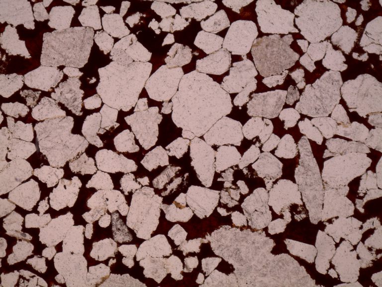
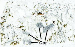
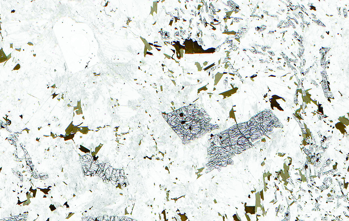
Fig 11.3.1 Corundum Granite
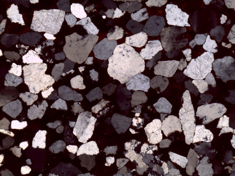
This is a rock from the former Bozeman Corundum Mine in Montana. It contains several large blocky gray (very) high-relief porphyroblasts of corundum and many smaller corundum grains. They are surrounded mostly by orthoclase and quartz. Some small flakes of greenish biotite are also present. As is common, the brittle corundum shattered somewhat during thin-section making. Some green-brown biotite is also present, as well as minor plagioclase. The corundum shows 1st-order interference colors and contains small included grains of deep-red rutile that appear nearly opaque in the XP view. Photos from www.rockptx.com. FOV = 25 mm.
This rock, of unknown origin, contains many high-relief blocky corundum crystals. The corundum shows up to 1st-order yellow and orange interference colors. Some of the corundum grains are shattered. These photos are from alexstreckeisen.it. FOV = 7 mm.
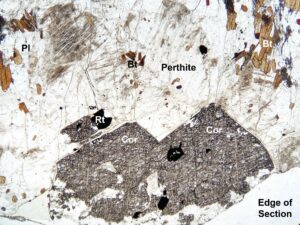
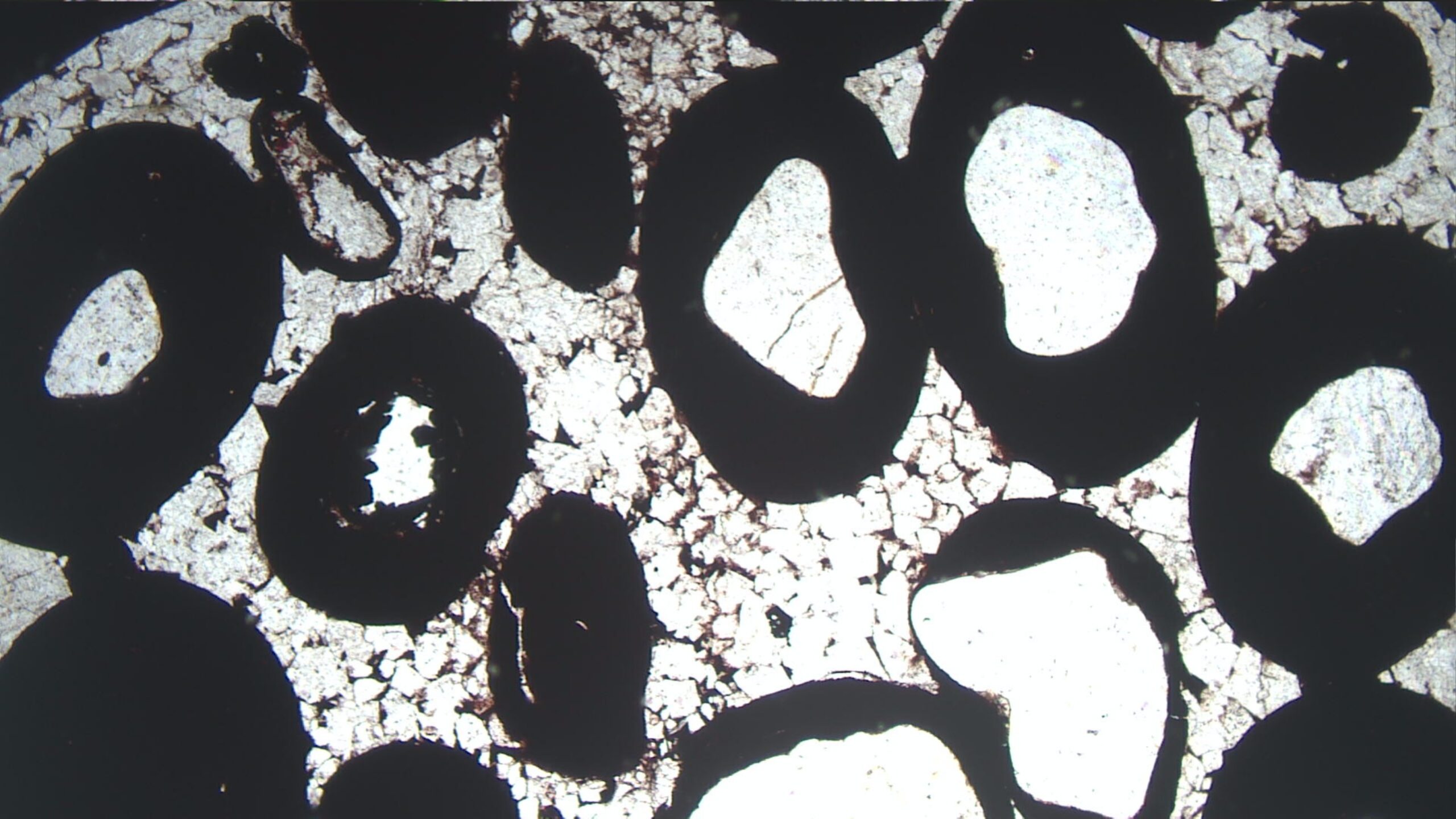
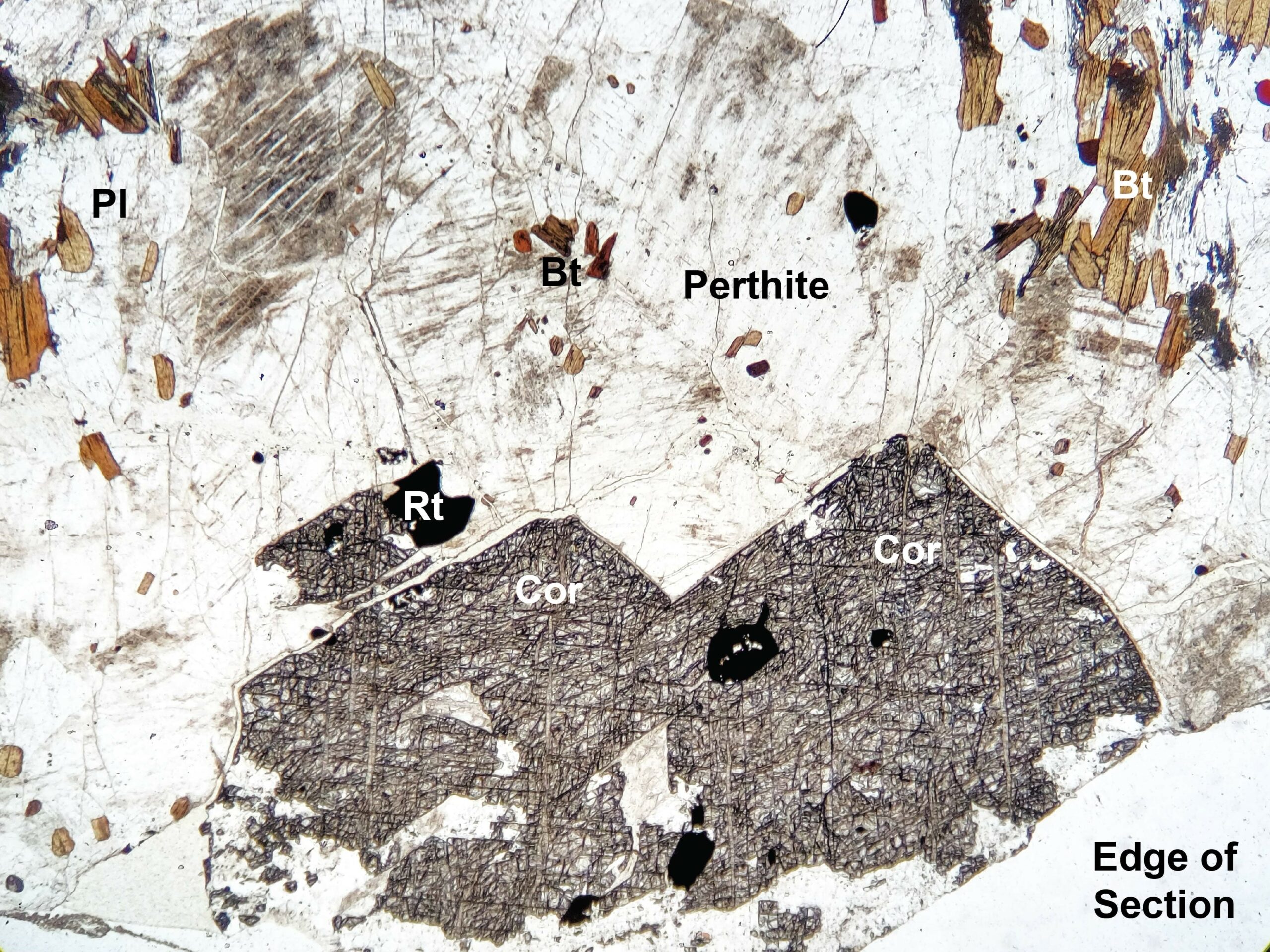
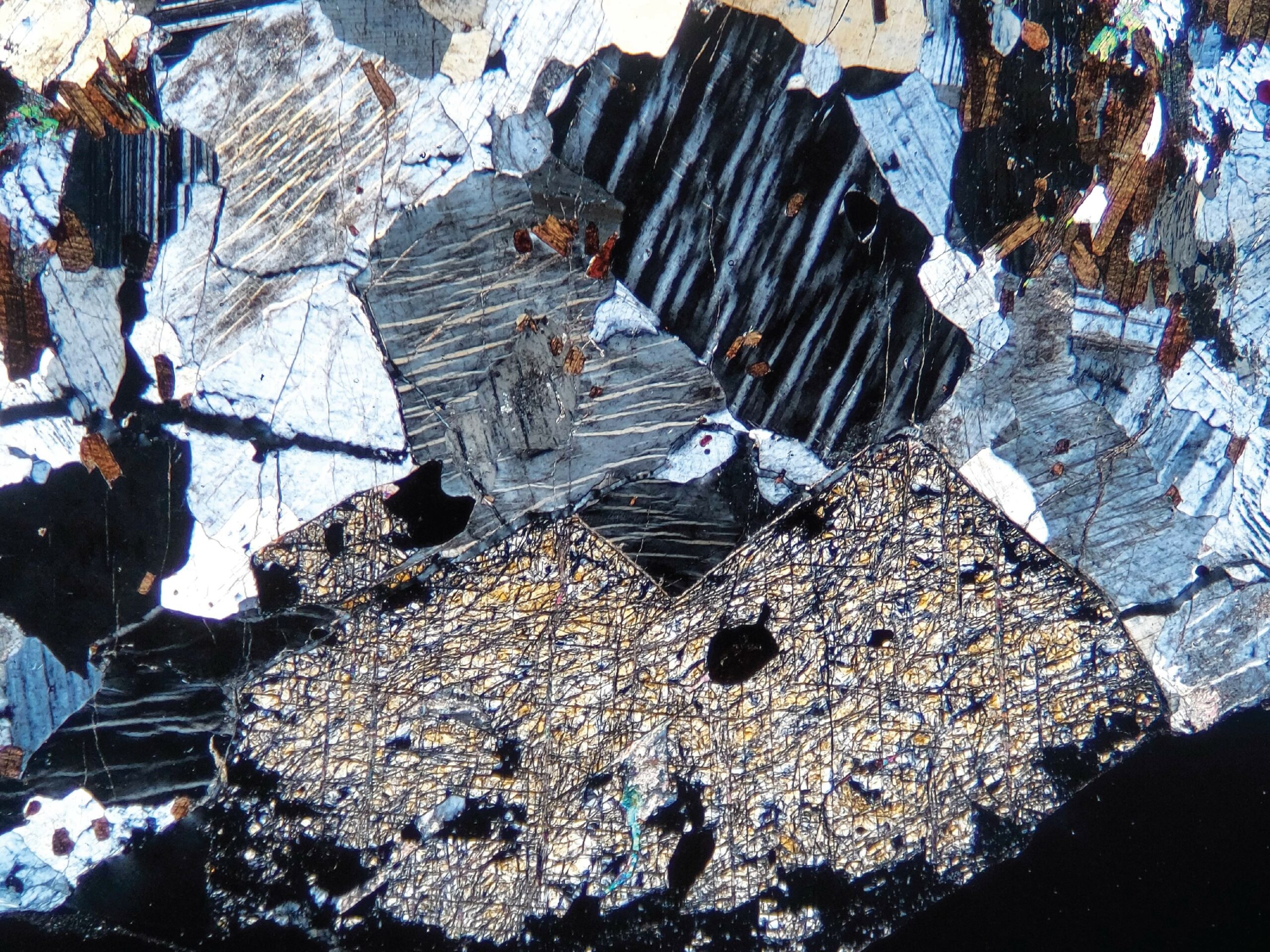
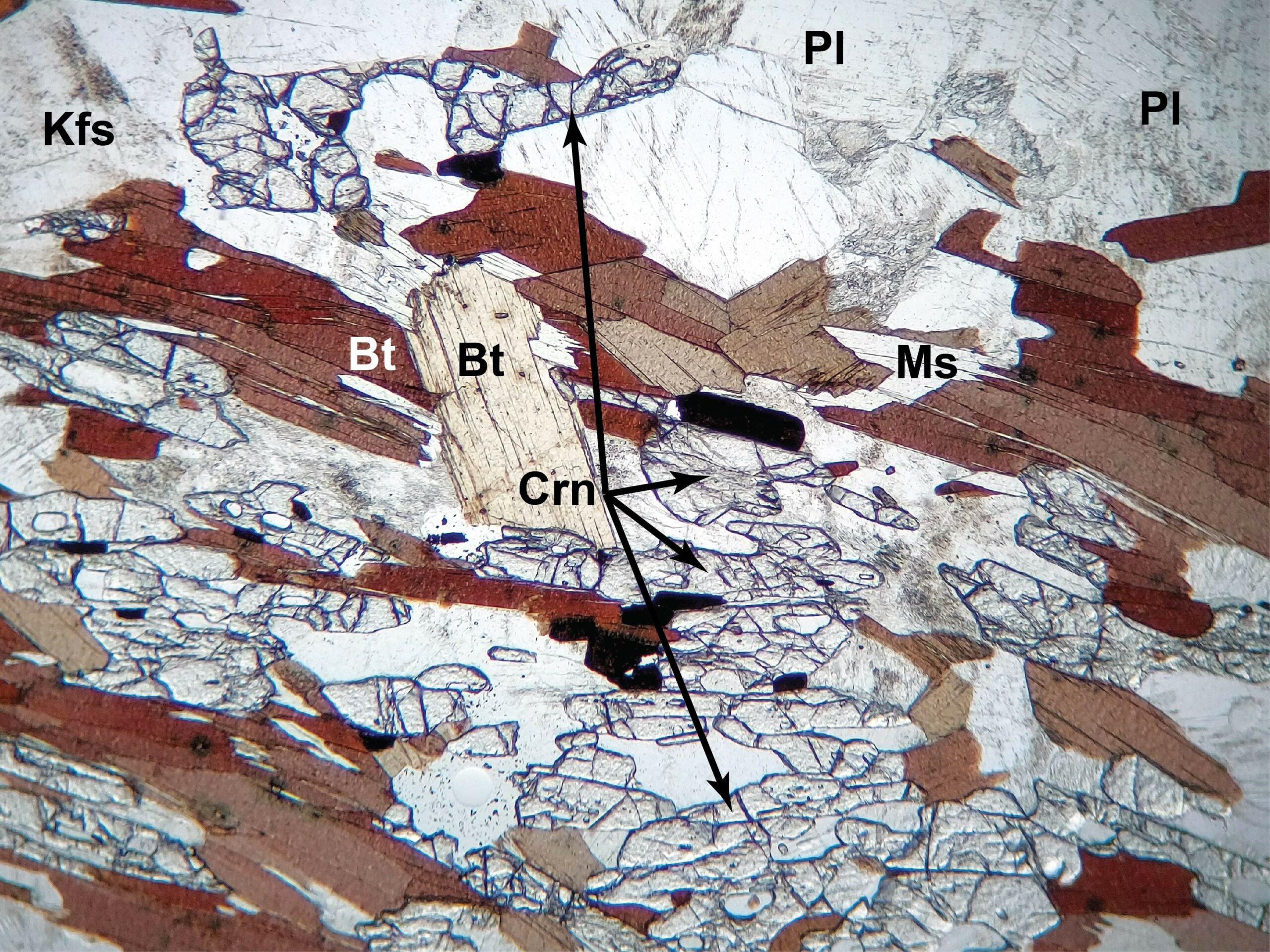
Corundum often occurs as chunky crystals surrounded by coronas or mantles of either plagioclase or potassium feldspar. In this schist from Montana, coarse high-relief and colorless corundum is surrounded by low-relief colorless perthite (K-feldspar with plagioclase exsolution lamellae). The perthite is slightly altered to brownish clay (PP). Other minerals are reddish-brown biotite and rutile (which looks opaque in these images; PP). In XP, corundum shows typical lower 1st-order yellow-orange colors; perthite is 1st-order gray to white. Biotite’s strong color obscures its 2nd-order interference colors, but its birds eye mottling is distinctive. FOV = 8 mm.
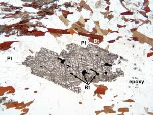
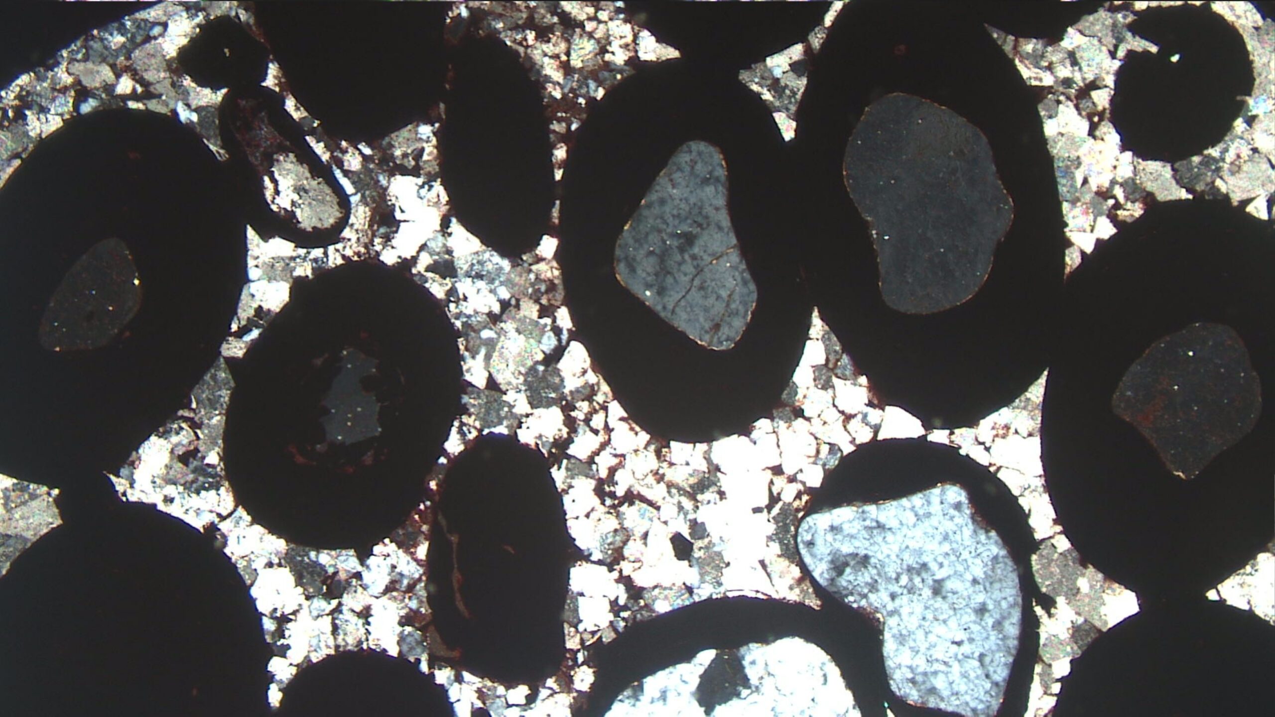
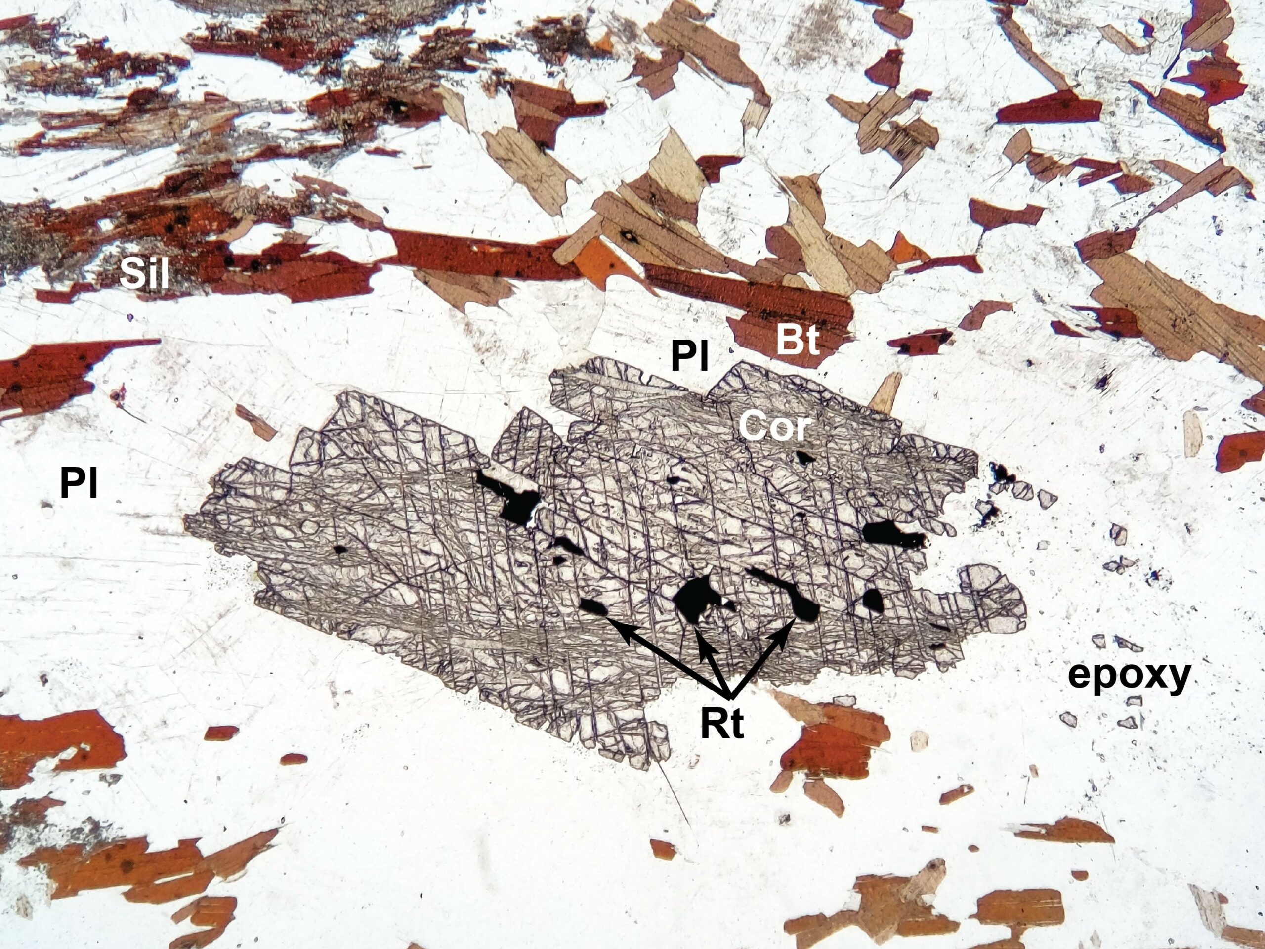
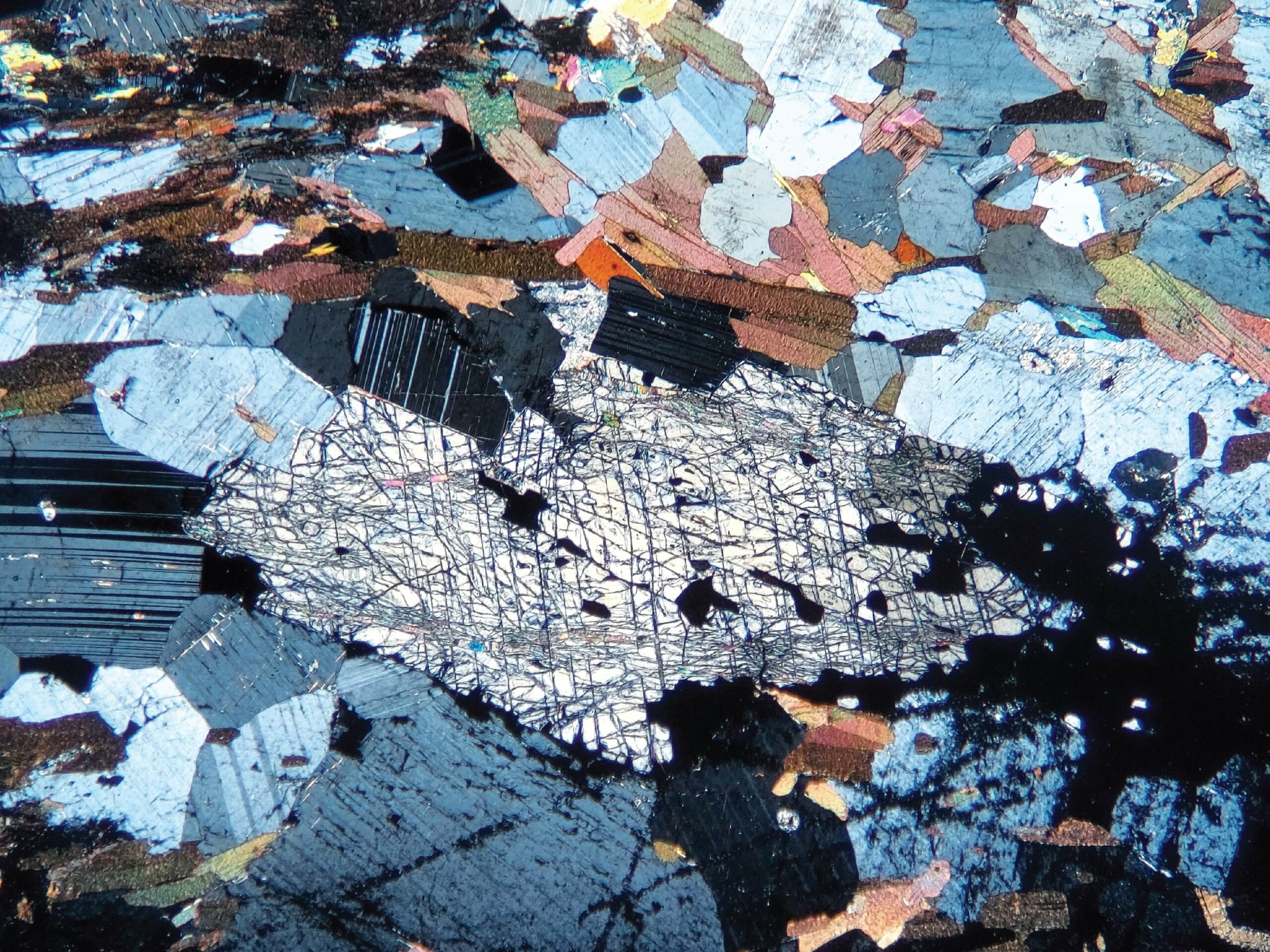
This schist from Montana contains large crystals of high-relief colorless corundum. In the PP view, we see that the corundum is surrounded by low-relief colorless plagioclase, reddish-brown pleochroic biotite, high-relief fine-grained sillimanite, and rutile that looks opaque (but is actually dark amber). In XP, corundum shows 1st-order yellow interference colors, plagioclase is 1st-order gray to white, biotite has 2nd order colors with well-developed birds eye mottling, and sillimanite is too fine-grained to make out its interference colors at this magnification. FOV = 8 mm.
This schist from Montana contains abundant colorless, high-relief corundum, together with colorless low-relief plagioclase and K-feldspar, brown biotite, and a bit of moderate-relief, colorless muscovite. Biotite shows intense normal pleochroism, and is much darker when cleavage planes run horizontally than when they are vertica. In XP, corundum in the center shows parallel extinction. Inclined grains towards the top and bottom of the image show characteristic 1st-order yellow to orange interference colors. K-feldspar and plagioclase have 1st-order gray to white interference colors, muscovite and biotite display 3rd-order colors. There is no quartz in this rock. FOV = 3.5 mm.
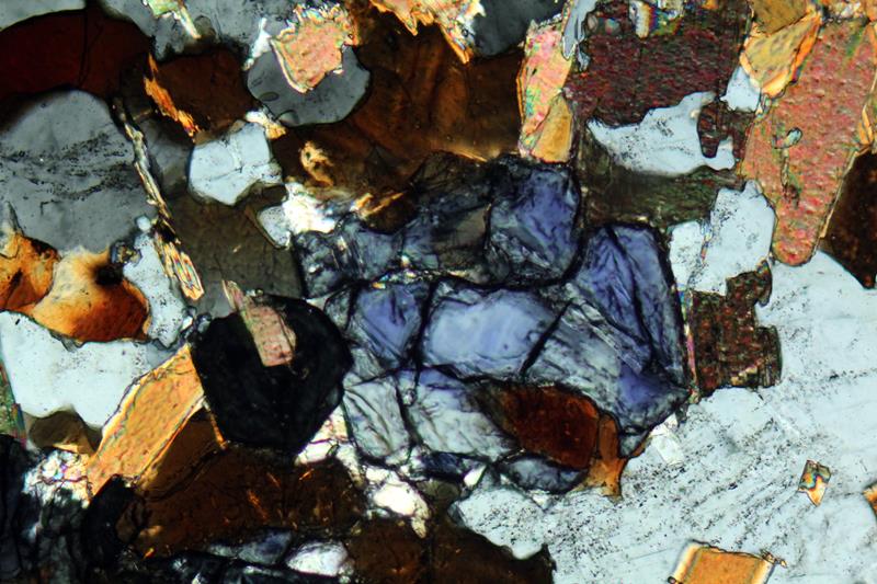
This zoned corundum (blocky and high-relief) contains blue (sapphire) cores. Brown biotite and colorless orthoclase are also present. In the XP view, the corundum shows anomalous blue interference colors. Biotite displays typical 2nd-order colors with birds-eye extinction, and the orthoclase shows only 1st-order gray colors. Photos from alexstreckeisen.it. FOV = 2 mm. Unfortunately, the PP and XP view are not perfectly aligned.
Occurrence—Spinel is the name of a cubic mineral with composition MgAl2O4. It is also the name of a mineral group with general composition AB2O4. The A atoms are most commonly Mg, Zn, Fe, or Mn. The B atoms are most commonly Al, Fe, or Cr. So, the spinel group includes many species, and the different species occur in different kinds of rocks. Solid solutions are common.
Some spinel-group minerals are metallic and opaque in thin section and will be considered separately. Here we focus on non-metallic spinels. MgAl2O4 (spinel) is by far the most common and important mineral of this spinel group. It is a high-temperature mineral found in marbles, metamorphosed marls, schists, or gneisses. It also occurs an accessory in mafic and ultramafic igneous rocks.
Distinguishing Features—Distinguishing the different kinds of spinels is sometimes challenging or impossible without chemical analysis. Spinel is cubic, so crystals are isotropic. They show very high relief and display no cleavage but may be fractured. Twinning is common. Euhedral to subhedral crystals often form equant grains and may display rhombic sections of octahedra. Spinel may also be interstitial (for example, Fig 11.4.2); filling irregular spaces between other grains in igneous rocks.
In general, crystals are colorless to green, light-red, brown, and less commonly other colors. End member MgAl2O4 spinel is colorless but becomes green (a variety called pleonaste) if Fe is present. Hercynite, end member FeAl2O4, is normally black. Spinel from mantle peridotites is typically reddish, brownish, or brownish-green. Pink and bluish varieties are also known.
Similar Minerals—Garnet and spinel are both isotropic and have high-relief, but spinel is almost always moderately to strongly colored and rarely contains inclusions. Garnet is typically lighter colored or colorless and commonly poikiloblastic. Spinel is sometimes difficult to distinguish from periclase, although periclase typically alters to brucite. Perovskite may resemble spinel but has a much higher refractive index.
spinel
Optical Properties ■■ Cubic; isotropic ■■ n = 1.72-1.74; relief is high ■■ Spinel display strong colors ■■ Isotropic character is a key identification
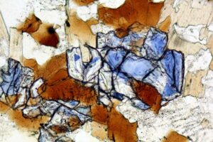
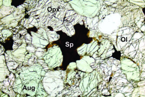
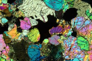
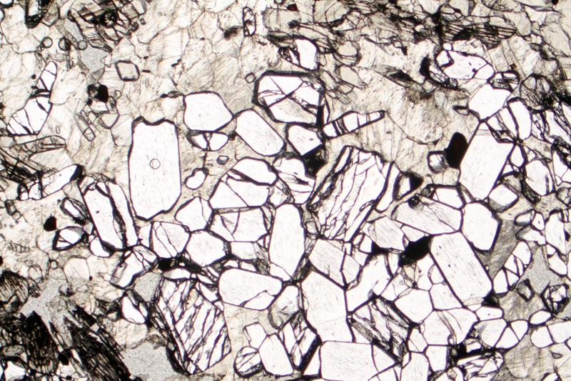
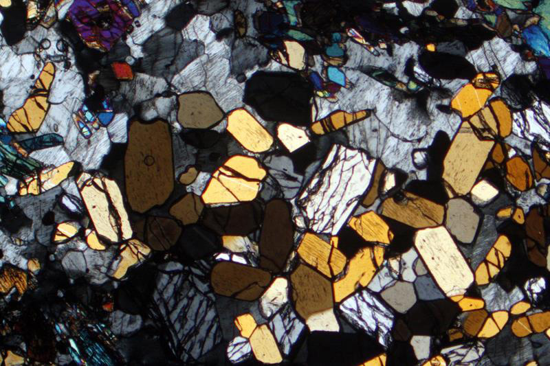
Fig 11.4.1 Spinel Peridotite
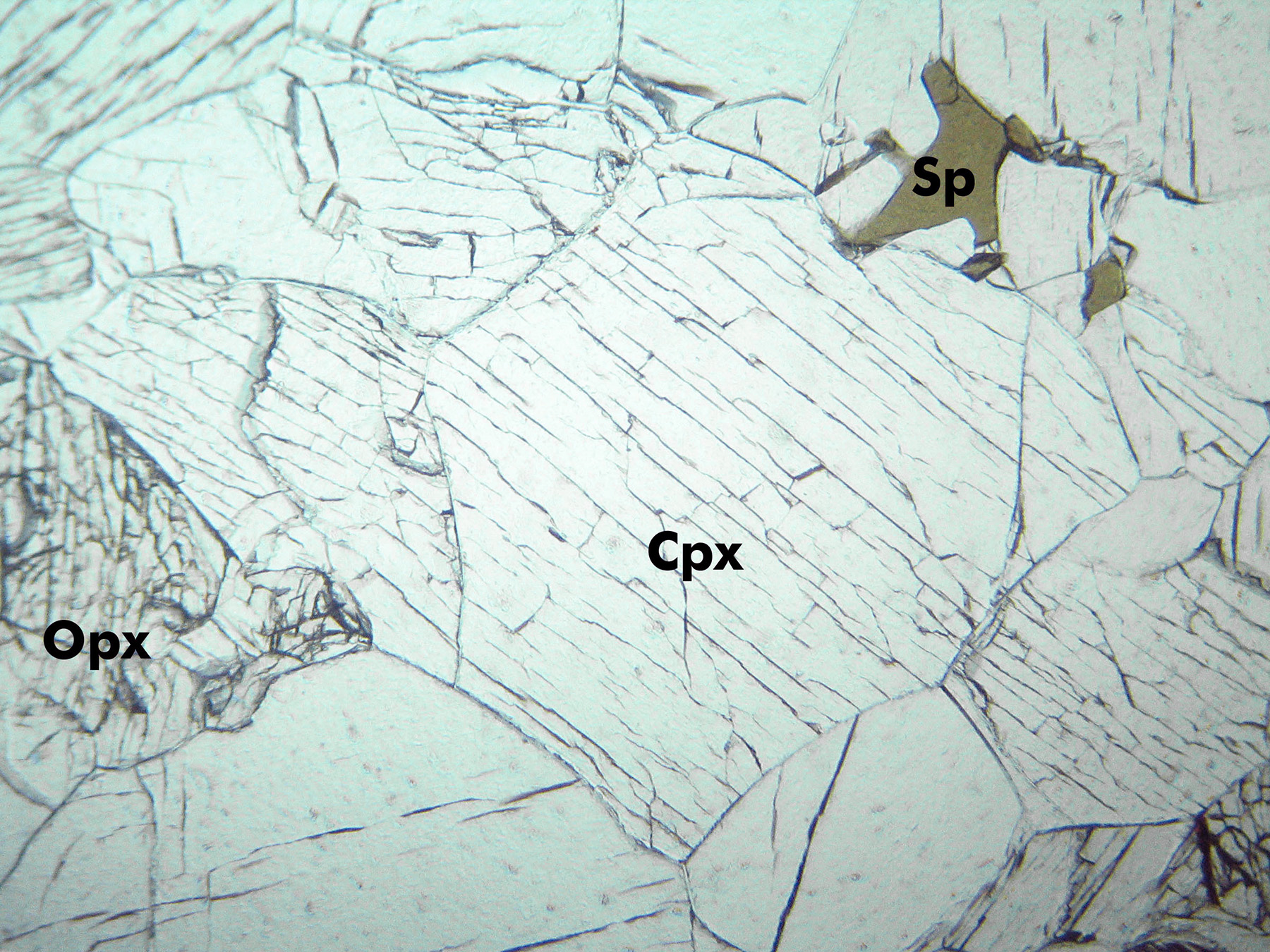
A high-relief green spinel grain occurs in the upper right (PP) in this peridotite xenolith from Kilbourne Hole, New Mexico. Most of the rest of the view is clinopyroxene (augite), except for a large grain of orthopyroxene, with slightly greater relief than the clinopyroxene, on the left edge of the photos. The orthpyroxene has typical 1st-order gray interference colors for that mineral; the augite has colorful higher-order colors. Spinel is isotropic – so appears black in the XP view. The field of view is 4.5 mm.
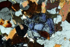
This spinel lherzolite contains orthopyroxene (colorless), olivine (colorless), augite (light green), and spinel (dark brown). In the XP view, the spinel is isotropic and the orthopyroxene shows only 1st-order gray interference colors. Augite displays 2nd-order colors of many hues, and the olivine shows higher-order colors. Olivine grains are more fractured than augite, and olivine’s interference colors are more blotchy. Photos from alexstrekeisen.it. FOV = 7 mm.
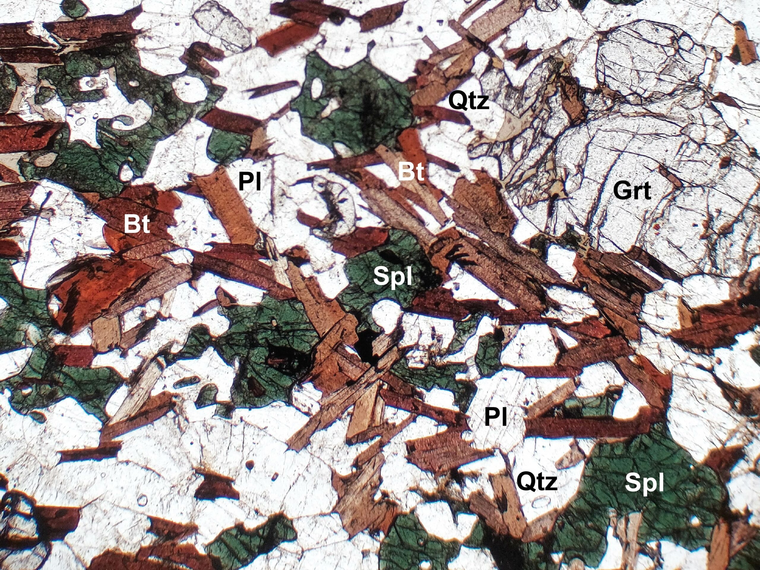
This photomicrograph shows a closeup view of a cluster of high-relief, equant, green spinel grains in a background of colorless plagioclase. The rock comes from Montana. In XP, a number of the spinel grains are not extinct, and still appear green. This is because the grains are small enough that they do not extend all the way through the thin section. If the light passes through a layer of plagioclase before reaching the spinel, the light is no longer plain polarized and the spinel grains simply add their color to it. FOV = 1.25 mm.
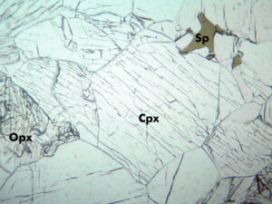
This wehrlite (olivine-clinopyroxene ultramafic rock) contains brown spinel (PP). Colorless clinopyroxene and olivine are hard to distinguish. In XP, the spinel is isotropic. The olivine shows 2nd-order colors; the augite displays only 1st-order interference colors Two elongate augite grains in the top-center part of the photo contain simple twins (XP). FOV = 9 mm. Photos from alexstrekeisen.it.
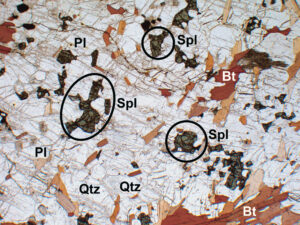
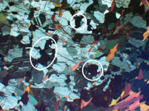
In this ultrahigh-temperature, granulite-facies metapelite from the North China Craton, high-relief green spinel (circled) dots a matrix of low-relief colorless quartz, plagioclase, and red-brown biotite (PP). High- temperature metamorphic spinel is commonly green, and high Ti makes biotite reddish. In XP, spinel is opaque, biotite shows 2nd- order colors, and quartz and plagioclase are 1st order gray and white. Sample courtesy of Dr. Shujuan Jiao, Chinese Academy of Sciences. FOV = 5 mm.
In this ultrahigh-temperature granulite-facies metapelite from the North China Craton, high-relief green spinel and red-brown biotite are surrounded by low-relief colorless quartz and plagioclase (PP). One irregular grain of istotropic garnet is also present. High- temperature metamorphic spinel is commonly green, and high Ti makes biotite reddish. In XP, spinel is opaque, biotite shows 2nd-order colors, garnet is black, and quartz and plagioclase are 1st-order gray and white. Sample courtesy of Dr. Shujuan Jiao, Chinese Academy of Sciences. FOV = 5 mm.
 Occurrence—Rutile is a common accessory mineral in metamorphic schists and gneisses. It also is found as small grains in intermediate to mafic igneous rocks. Less common occurrences are in veins, in pegmatites, and in some sediments.
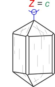


 Occurrence—Corundum can be found as an accessory mineral in metamorphosed carbonates, in highly aluminous metasediments, in some Al-rich igneous rocks, and in placers. Massive corundum is found in skarns.
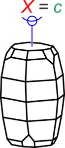


 Occurrence—Spinel is the name of a cubic mineral with composition MgAl2O4. It is also the name of a mineral group with general composition AB2O4. The A atoms are most commonly Mg, Zn, Fe, or Mn. The B atoms are most commonly Al, Fe, or Cr. So, the spinel group includes many species, and the different species occur in different kinds of rocks. Solid solutions are common.


