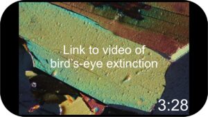
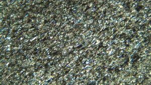
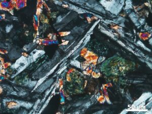
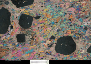
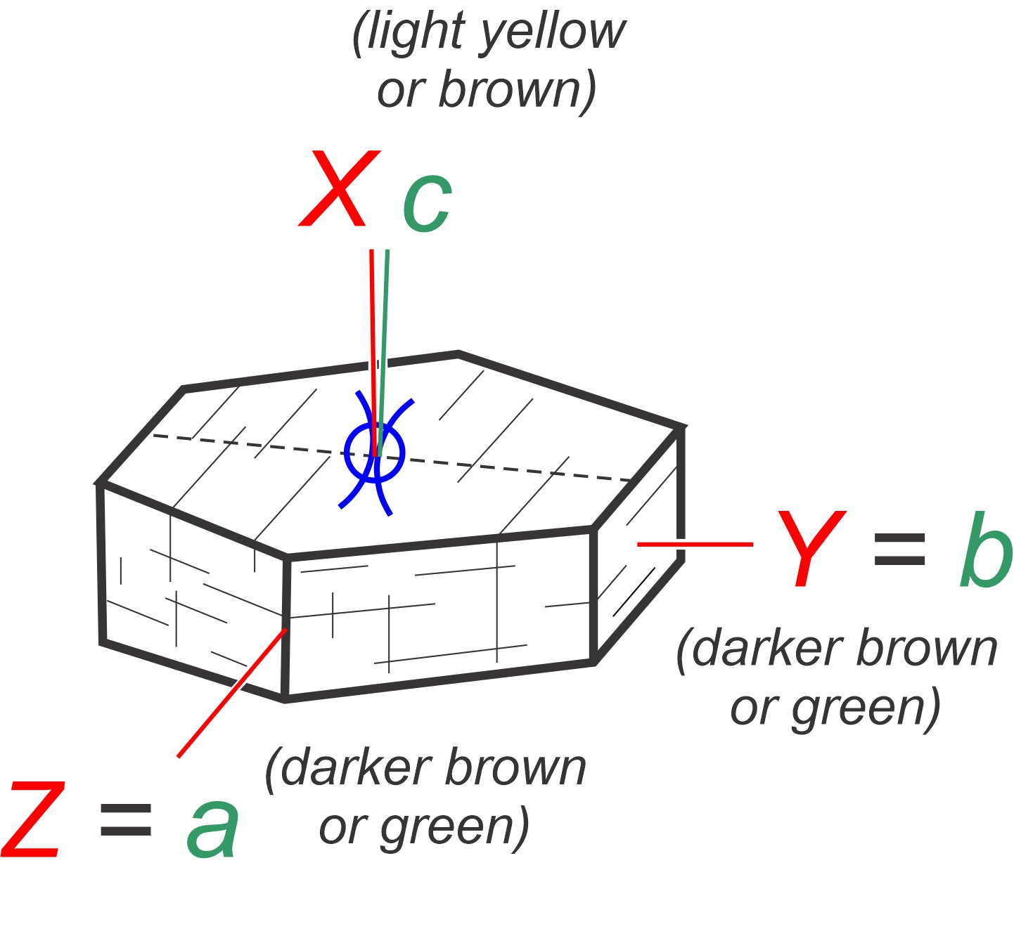
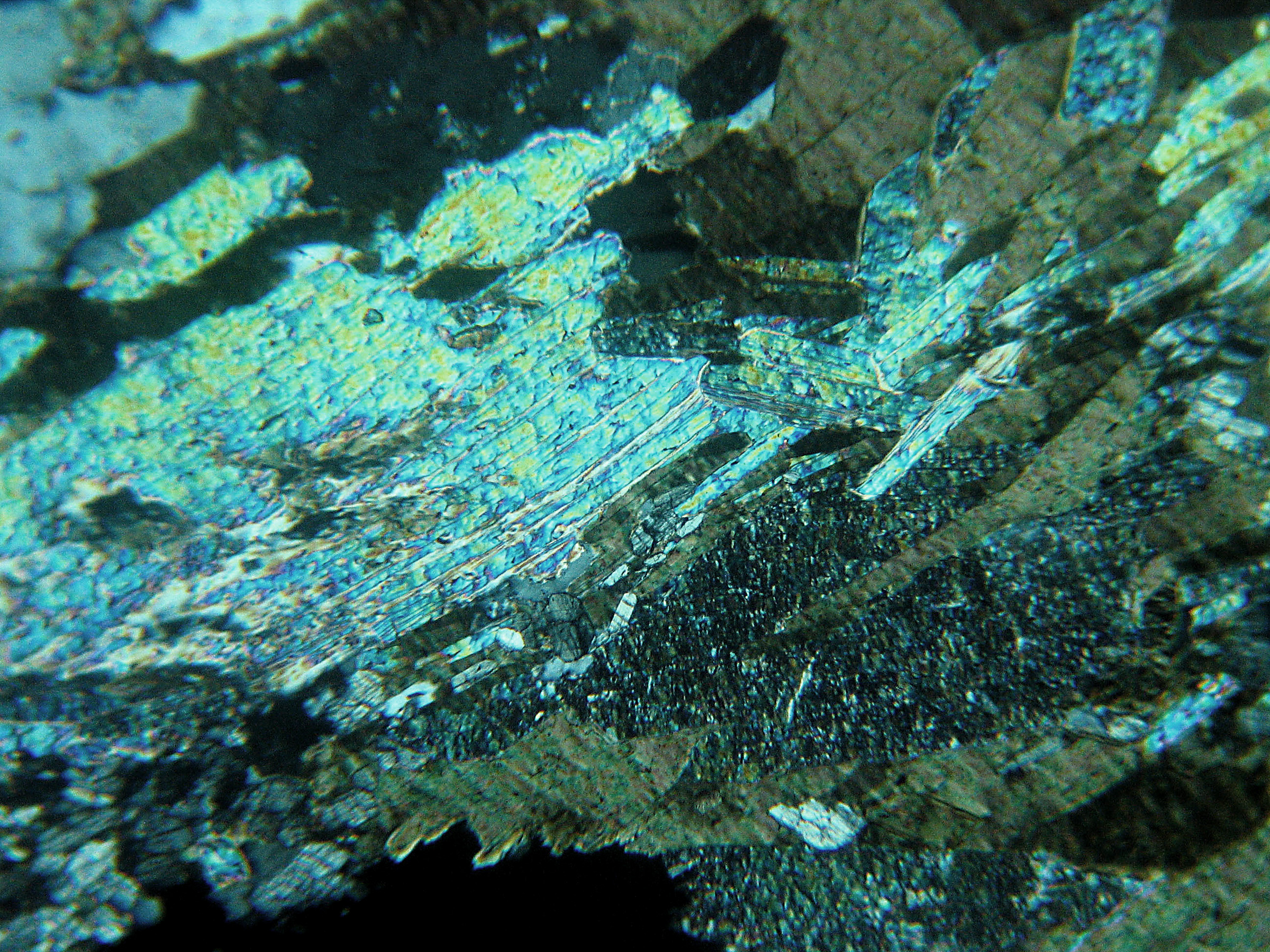
 Bird’s-Eye Extinction
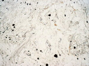
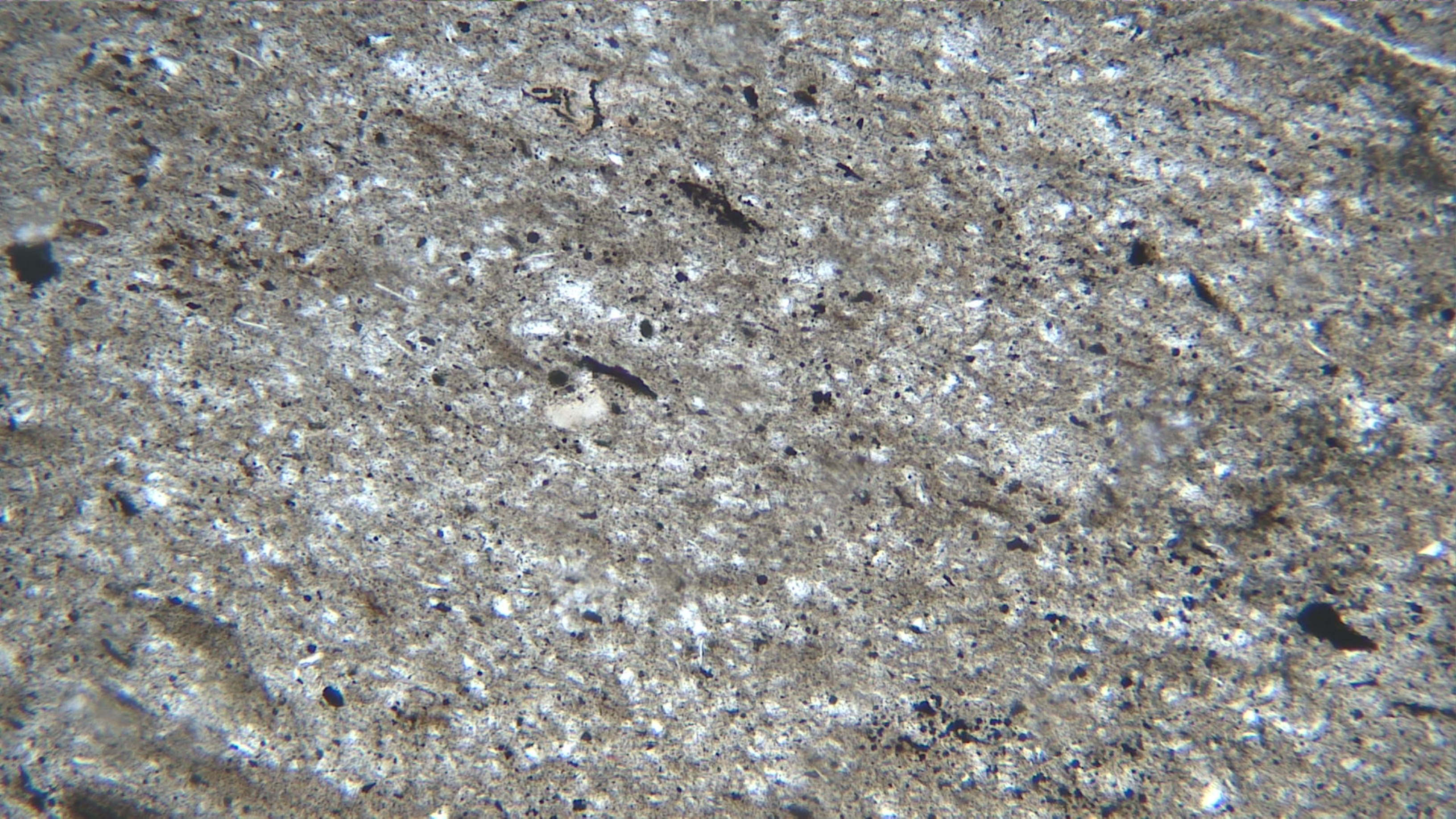
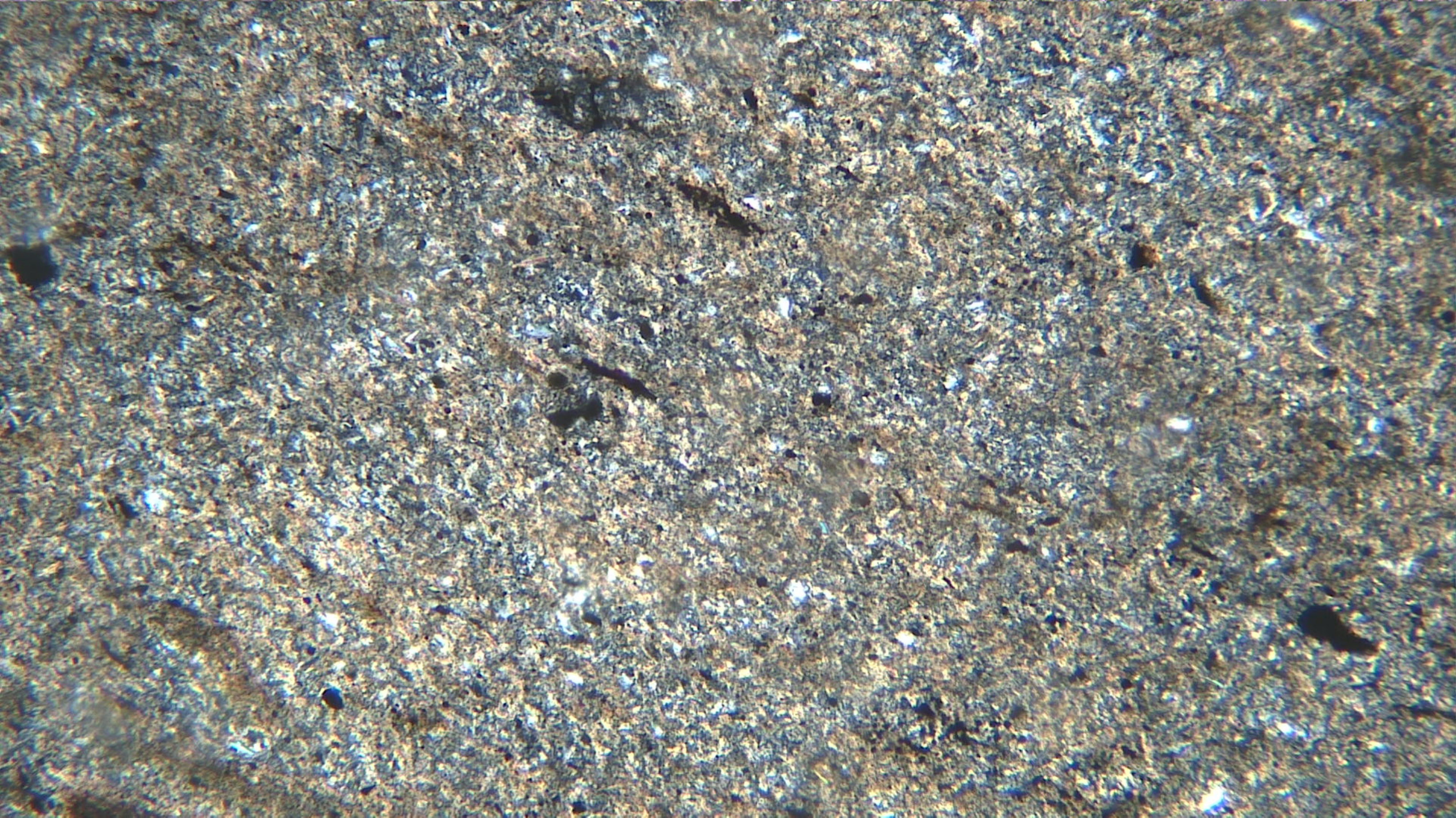
Some sheet silicates (notably biotite and muscovite, but also others) commonly show bird’s-eye extinction under crossed polars. It’s named after bird’s eye maple, and appears as a pebbly texture, especially when a mineral grain nears extinction. This video discusses bird’s-eye extinction and how it can be used to help identify minerals in thin section.
3.1 Serpentine
Mg3Si2O5(OH)4
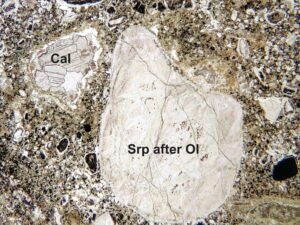
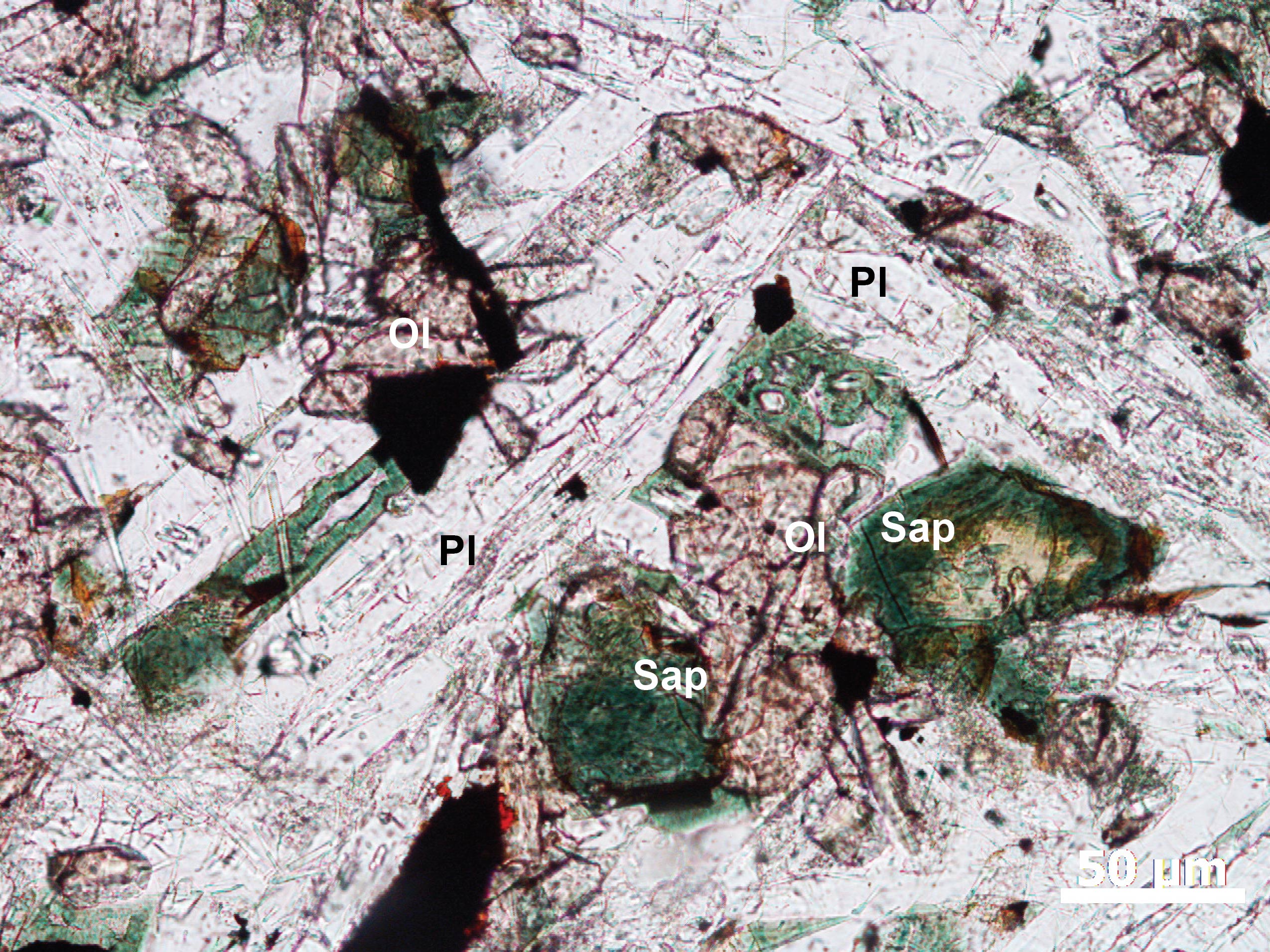
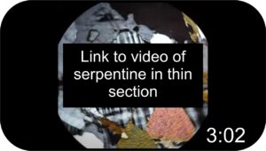 Occurrence—Serpentine is most commonly a secondary mineral in mafic and ultramafic igneous rocks, generally forming by alteration of olivine or pyroxene. It is typically associated with fine-grained magnetite or chromite (see for example Figure 3.1.2). Serpentine is also found in marbles, associated with carbonates, forsterite, dolomite, or magnesite. It may occur as replacements for individual grains, in massive aggregates, or in veins. A rock that consists mostly of serpentine is a serpentinite (which is the state rock of California).
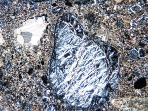
Distinguishing Features—Serpentine has three polymorphs: antigorite, lizardite, and chrysotile. They cannot be definitively distinguished without crystallographic or spectroscopic analysis, but different forms can sometimes have slightly different properties. Antigorite is micaceous or flaky, and is commonly in foliated or scaly masses. Lizardite is finer grained and may display a net-like pattern and undulatory extinction. Chrysotile is especially distinctive as fibrous masses in veins or mats. Antigorite has one perfect cleavage. Cleavage is not visible for other serpentine minerals.
Serpentine is generally colorless to green. It may be slightly pleochroic from clear to pale green to yellow green. It has low birefringence (maximum interference colors are 1st-order yellow).
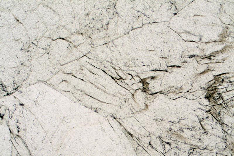
Serpentine is normally an alteration product, often as pseudomorphs after mafic minerals. It typically appears as an aggregate of small anhedral crystals and may exhibit a coarse “alligator skin” texture (Fig 3.1.4).
Similar Minerals—Chlorite is typically biaxial (+), more pleochroic, and displays lower or anomalous interference colors. Micas have much higher birefringence, and muscovite is generally not pleochroic, whereas biotite is much more strongly colored. Brucite is uniaxial, rare, and displays anomalous interference colors. Fibrous amphiboles have higher refractive indices and birefringence than chrysotile.
Optical Properties
■■ Monoclinic; biaxial (-) or (+)
■■ 2V variable
■■ α = 1.50-1.55, β = 1.50-1.60, γ= 1.50-1.60
■■ δ = 0.006-0.014, maximum interference colors are normally 1st-order gray or white, more rarely 1st-order yellow.
■■ Interference colors may be somewhat anomalous if crystals have a strong green color
■■ Interference figures are often hard to obtain due to fine grain size
■■ Extinction is parallel to cleavage
■■ Length slow
|
 |
|
|
|
|
|
|
|
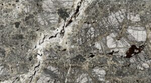
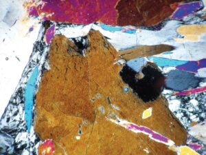
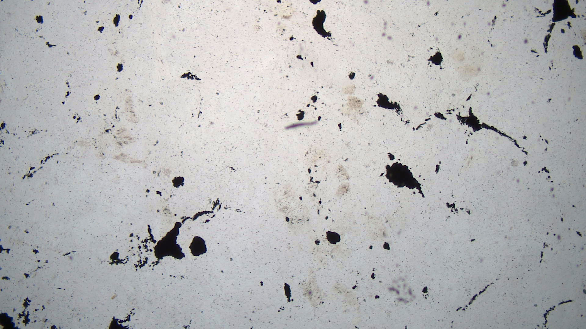
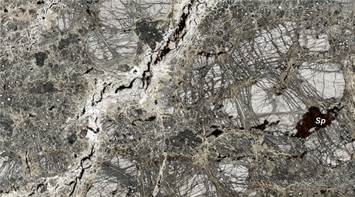
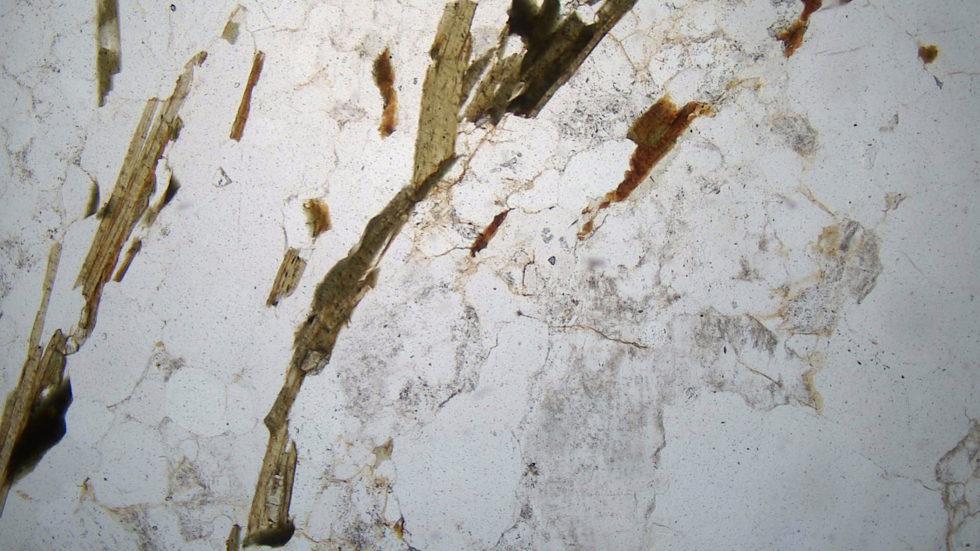
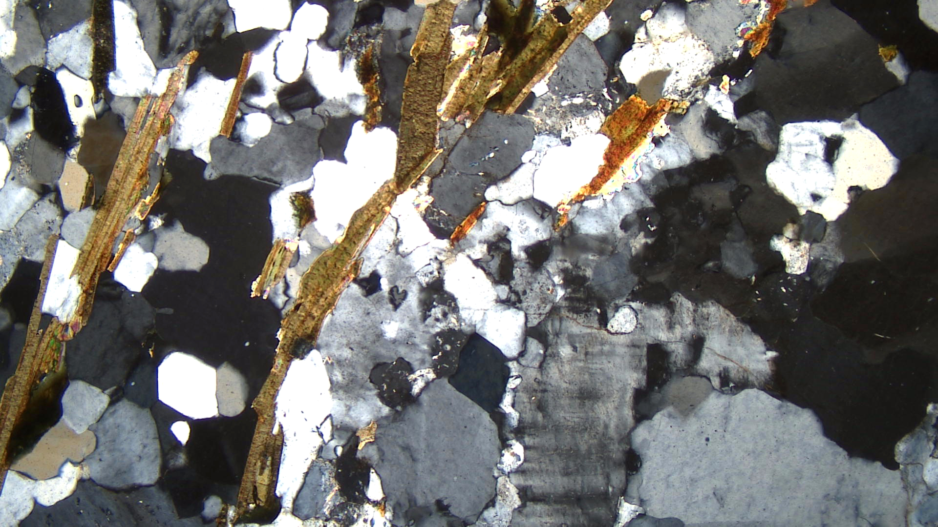
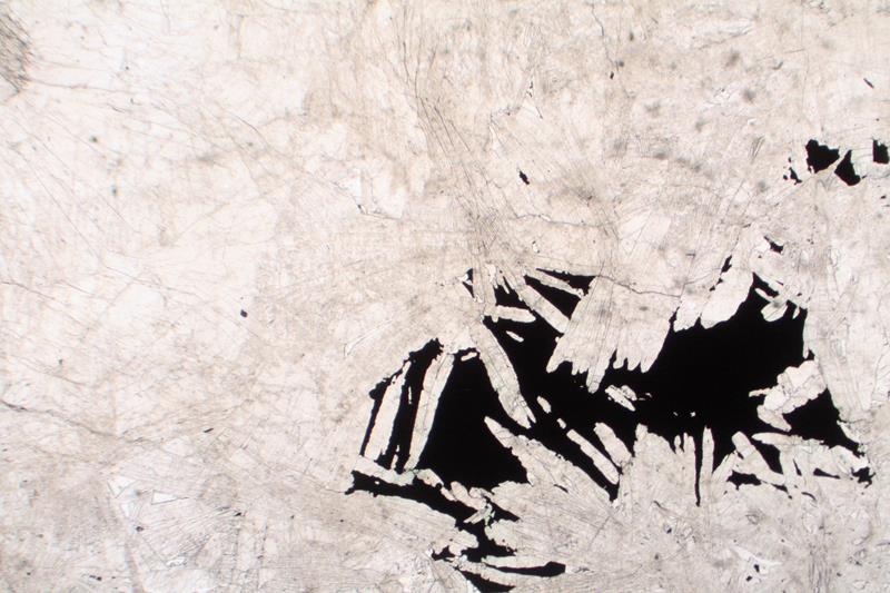
| Fig 3.1.5 Altered Peridotite
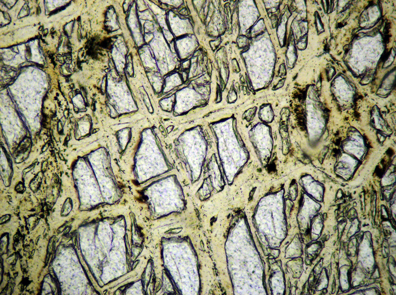
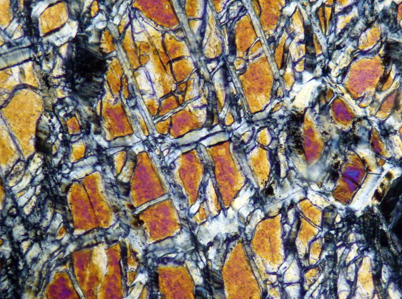
In this highly altered peridotite, serpentine is fine-grained greenish-gray in veins and patches (PP). The colorless grains are highly cracked pyroxene and possibly olivine. Several grains of dark-brown (almost black) spinel are present; the largest is labeled Sp. Olivine shows only 1st-order gray interference colors and the pyroxene grains show up to 1st-order orange colors (XP). The spinel is isotropic. FOV = 15 mm. Photos modified from virtualmicroscope.org. |
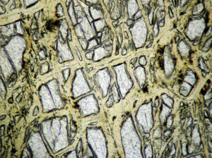
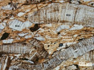
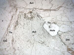
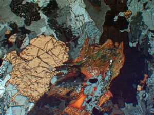
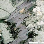
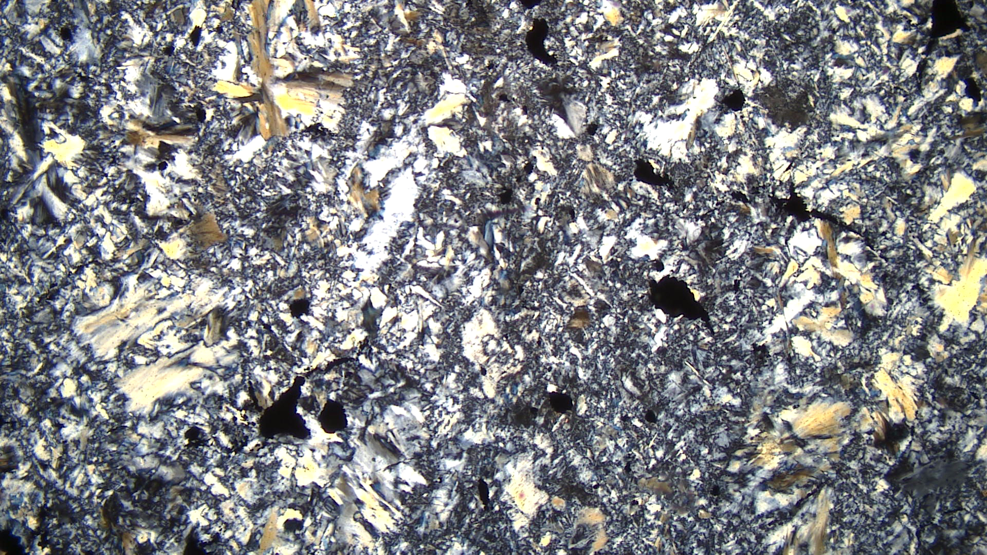
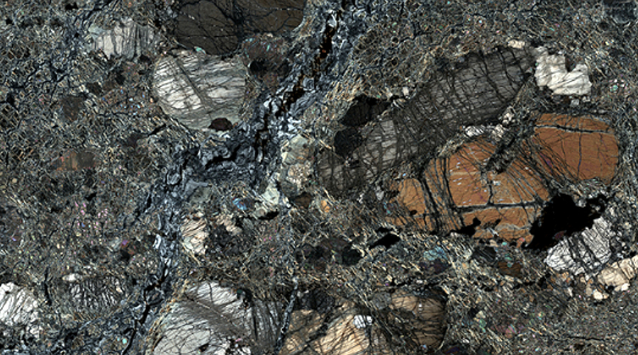
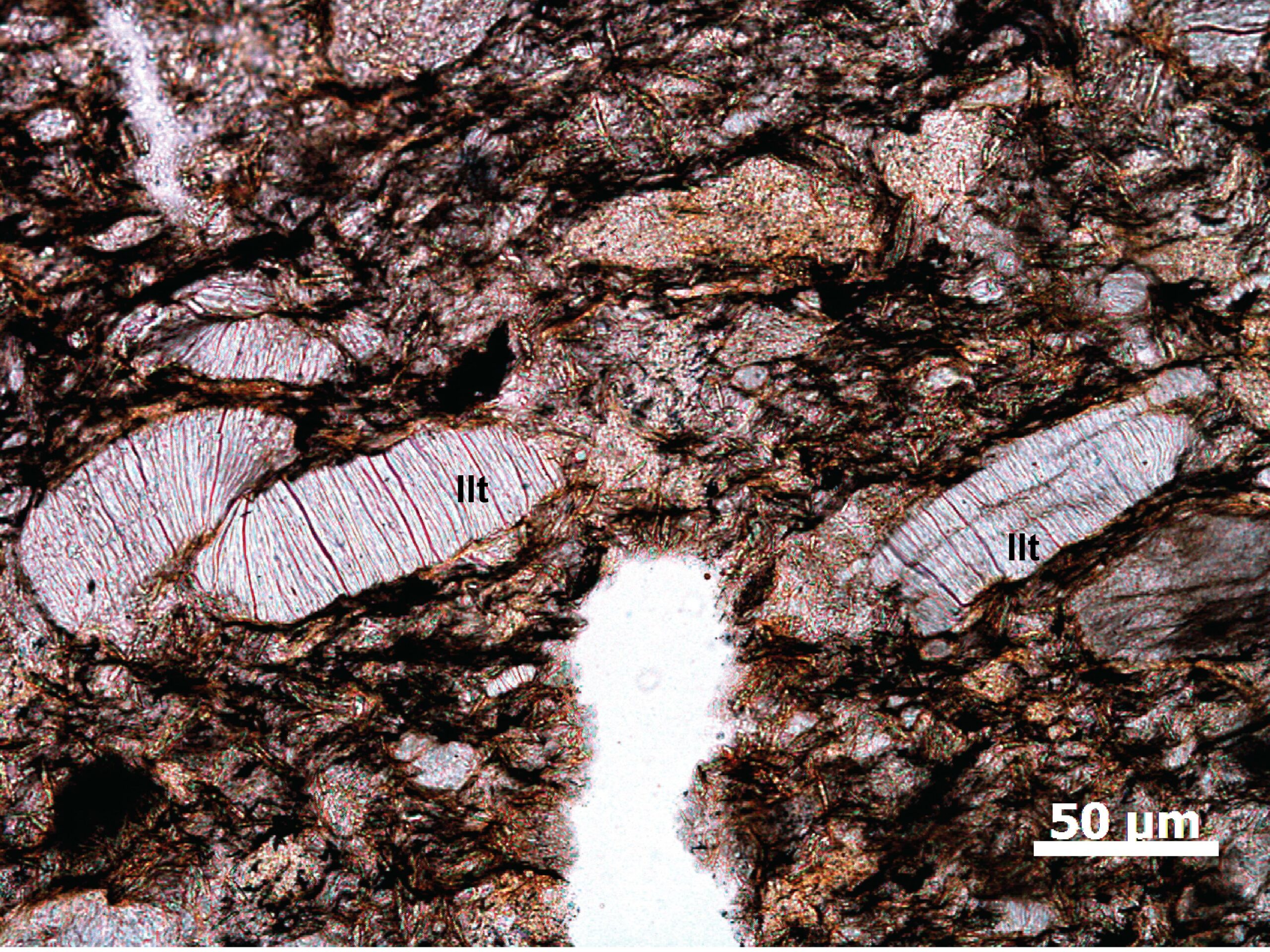
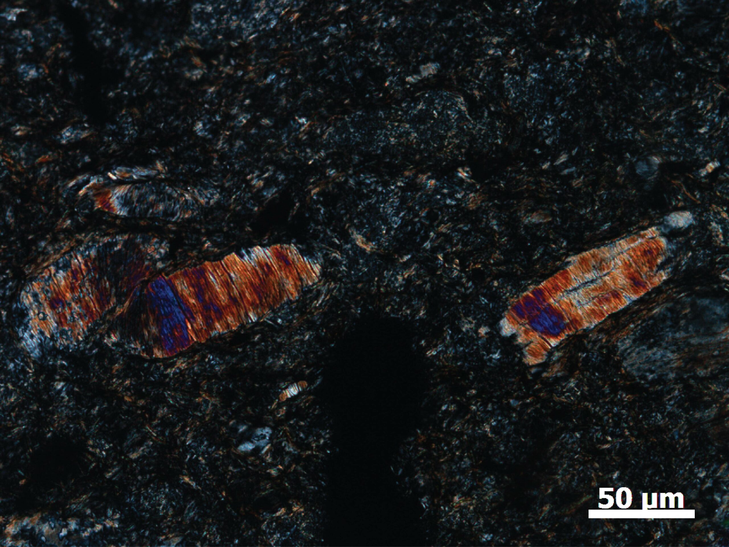
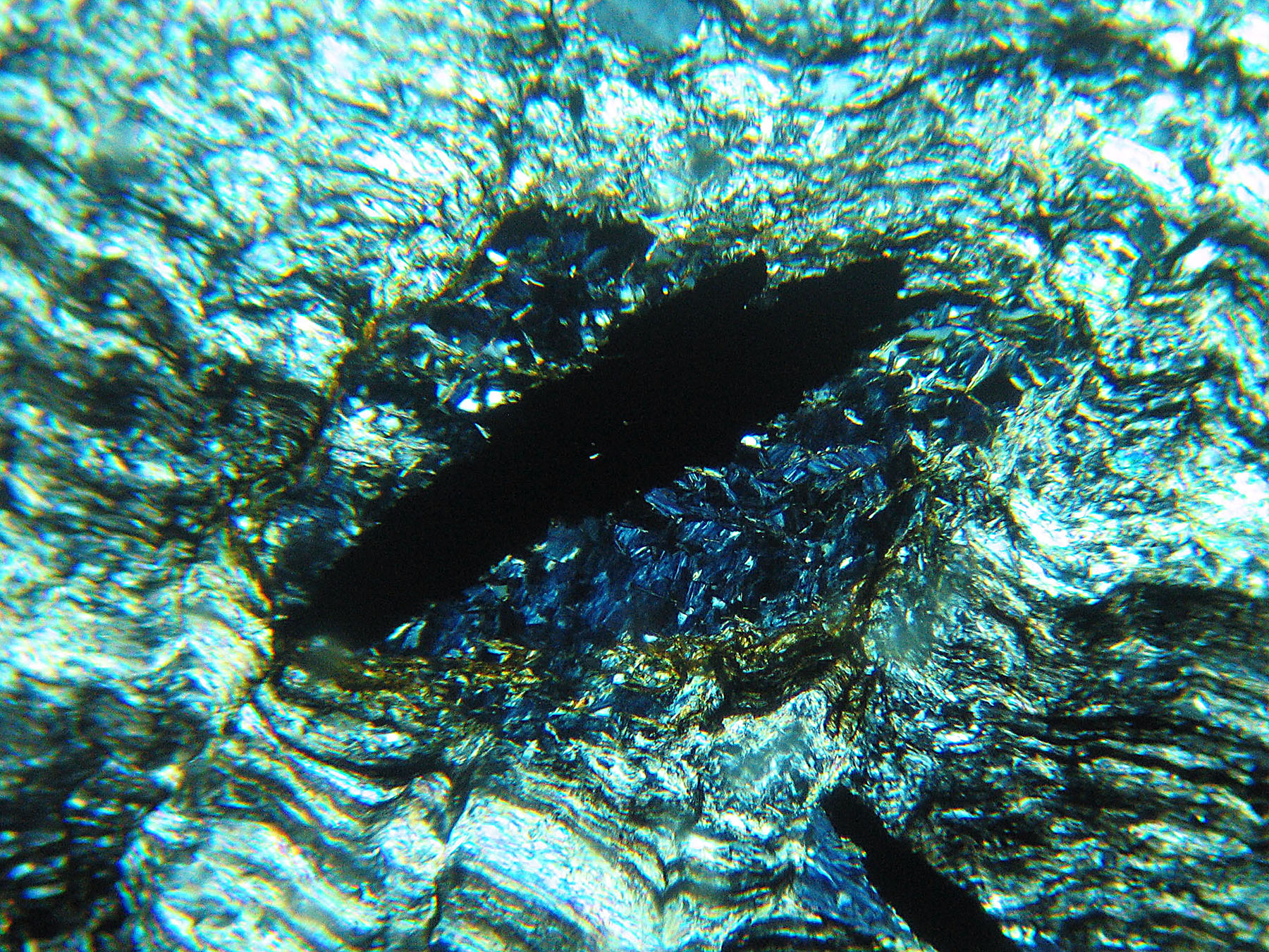
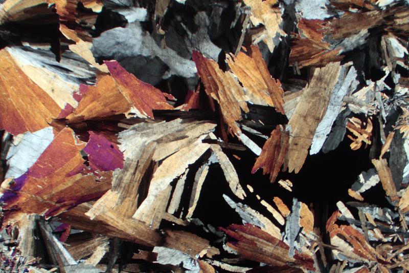
Fig 3.1.6 Altered Harzburgite
Antigorite in this metamorphosed harzburgite (opx- and ol-rich peridotite), is platy, unlike fibrous chrysotile, colorless to pale green, and in some ways resembles chlorite. Like chlorite, antigorite can have anomalous Berlin blue or brown interference colors, but, unlike chlorite, both colors can appear in the same rock at different orientations. Both can be seen in this thin section. The interference colors for chlorite, in contrast, depend on composition – almost entirely anomalous blue (Fe-rich), anomalous brown (Mg-rich), or anomalous violet (intermediate composition), irrespective of orientation. Photos from Dr. Kurt Hollocher. FOV = 1.2 mm. |
|
|
|
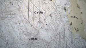
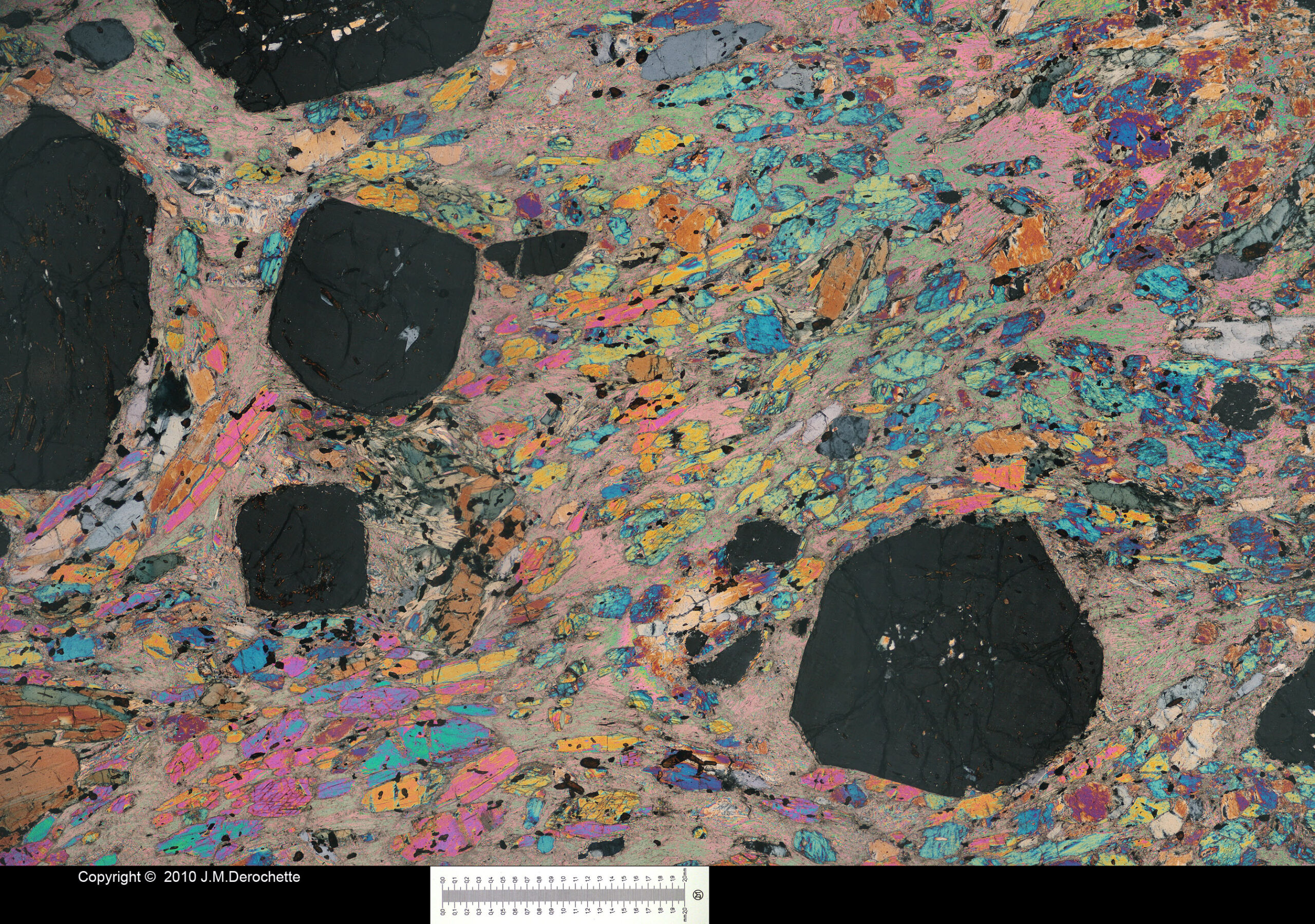
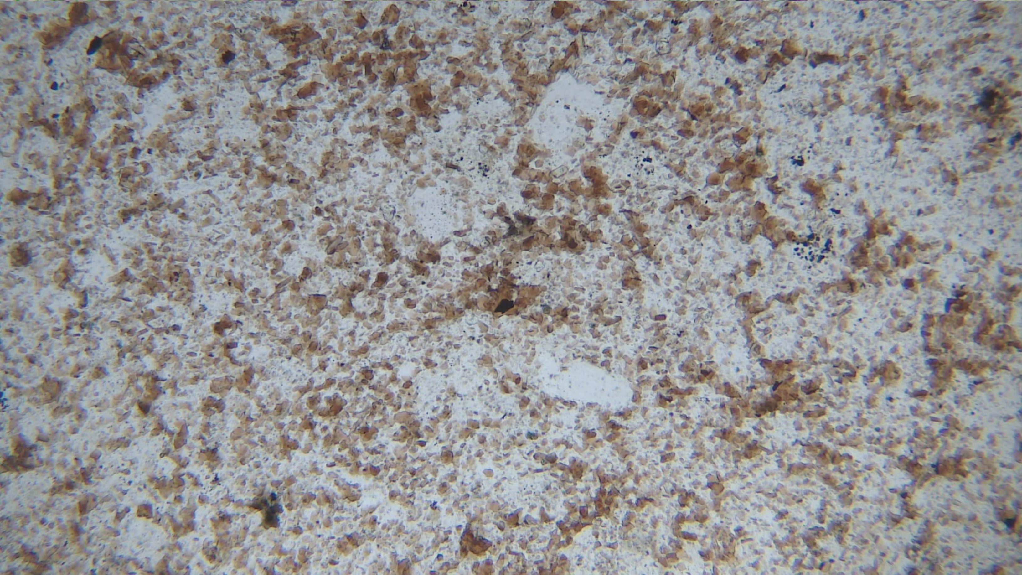
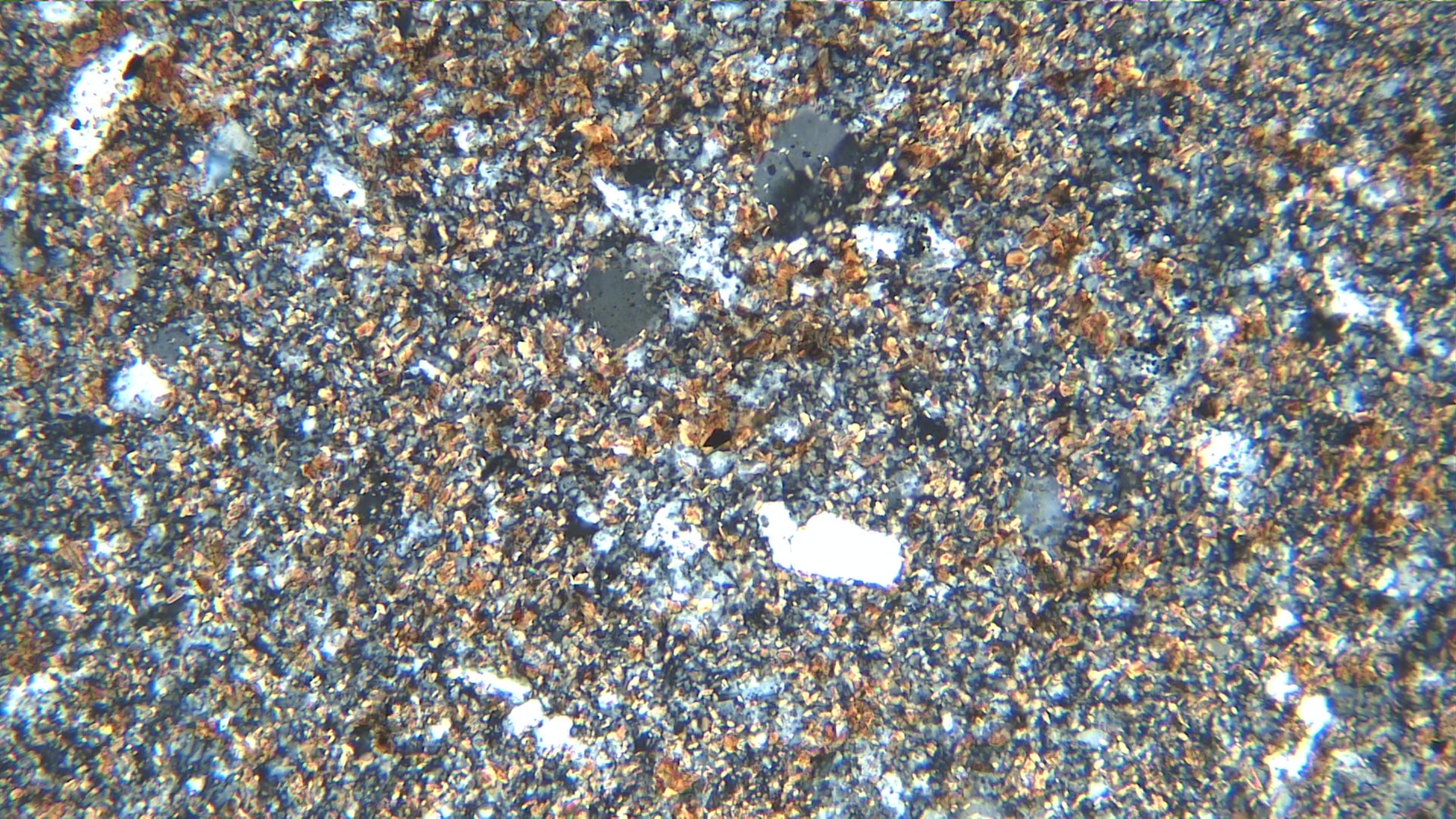
| Fig 3.1.7 Kimberlite
This altered kimberlite from Colorado contains abundant low-relief brownish serpentine and variable-relief brownish calcite, both as fine grains in the matrix and as coarse clots (PP). Large masses of serpentine pseudomorph olivine. Fine-grained brown biotite, too small to discern in this view, lends color to the matrix (PP). In XP, serpentine has characteristic 1st- order gray interference colors and a veiny texture, and calcite is pearly. FOV = 8.5 mm. |
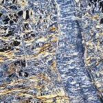
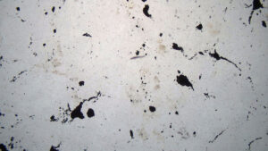
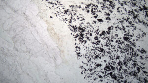
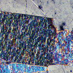
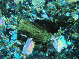
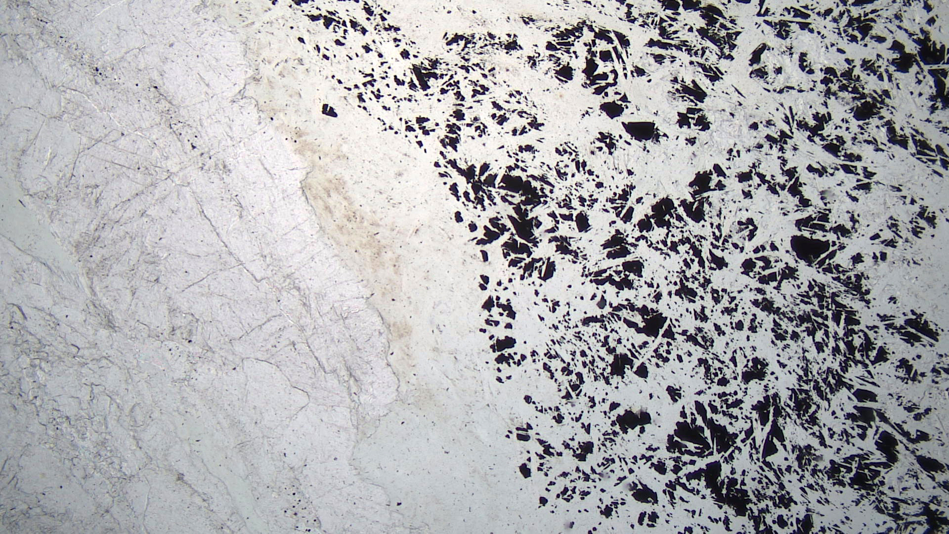
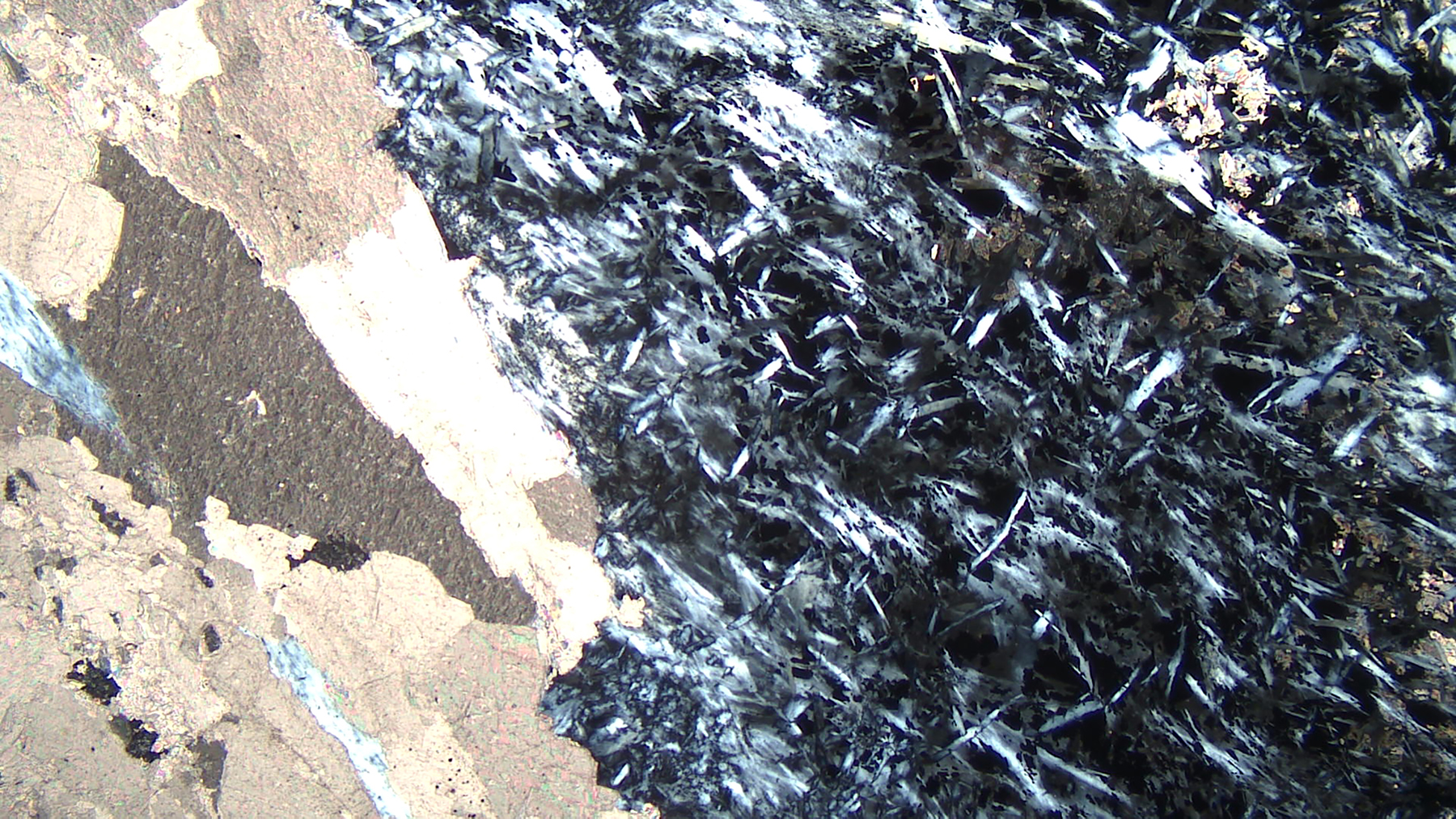
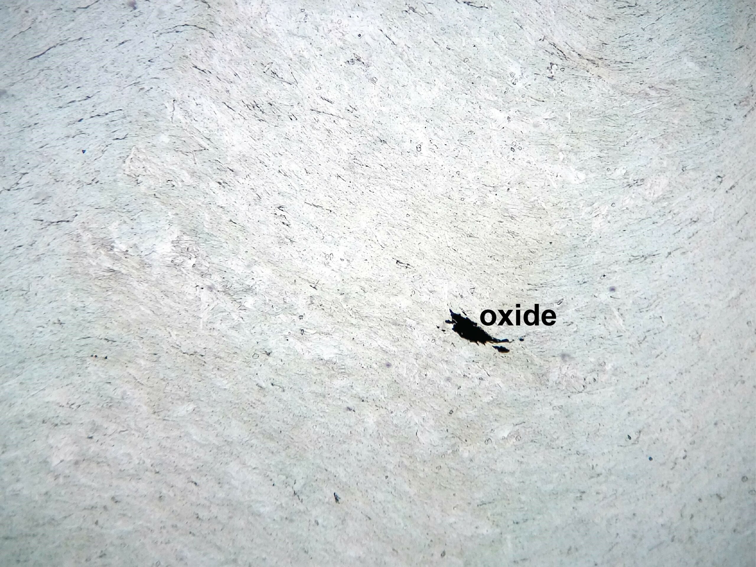
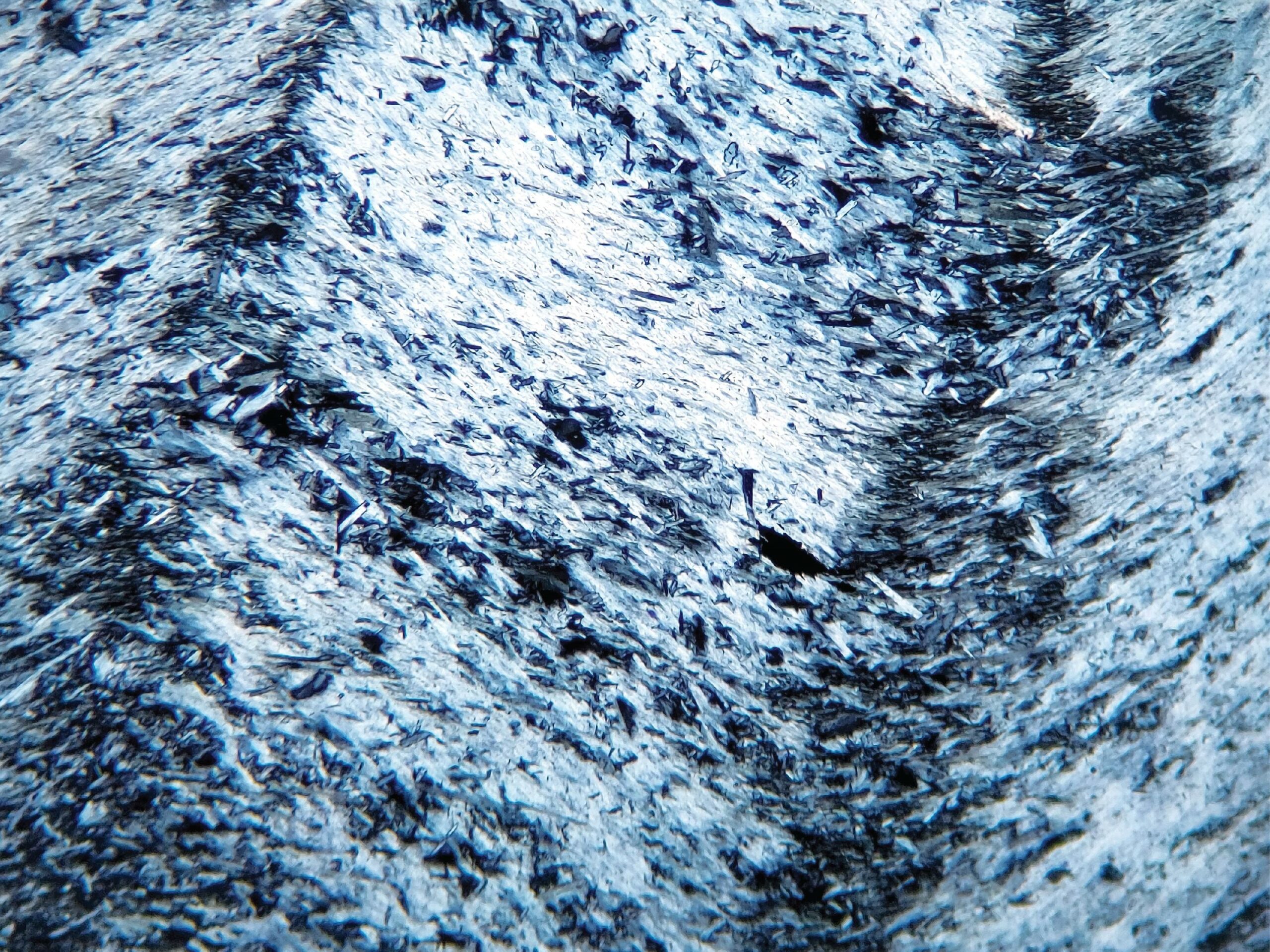
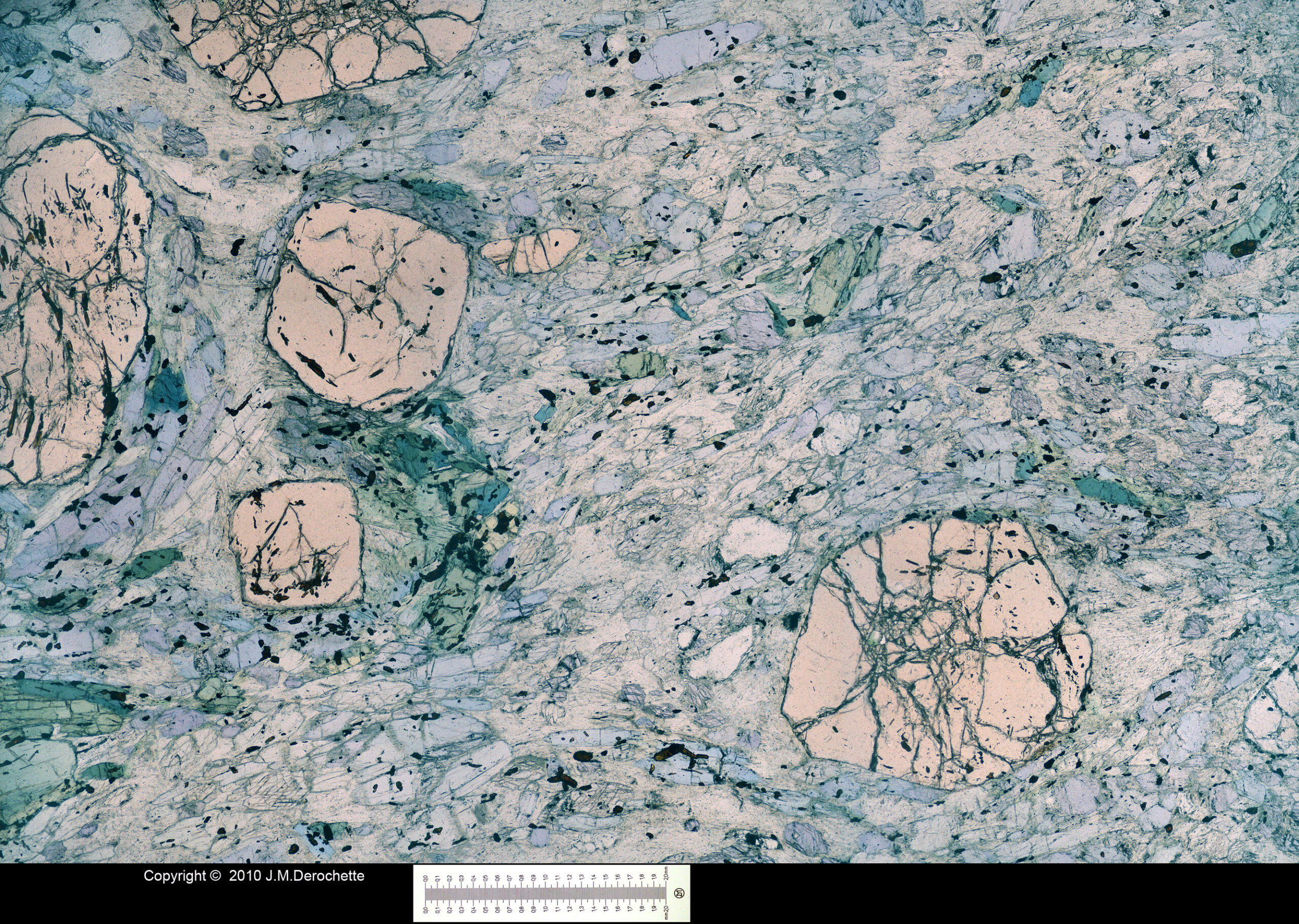
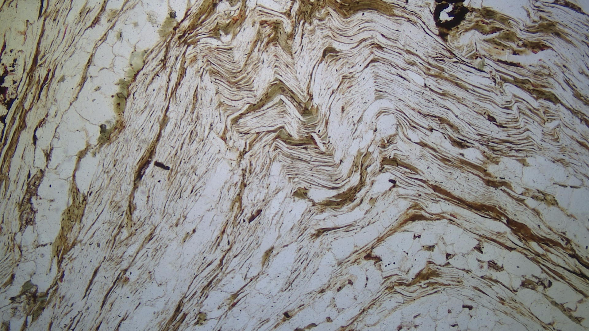
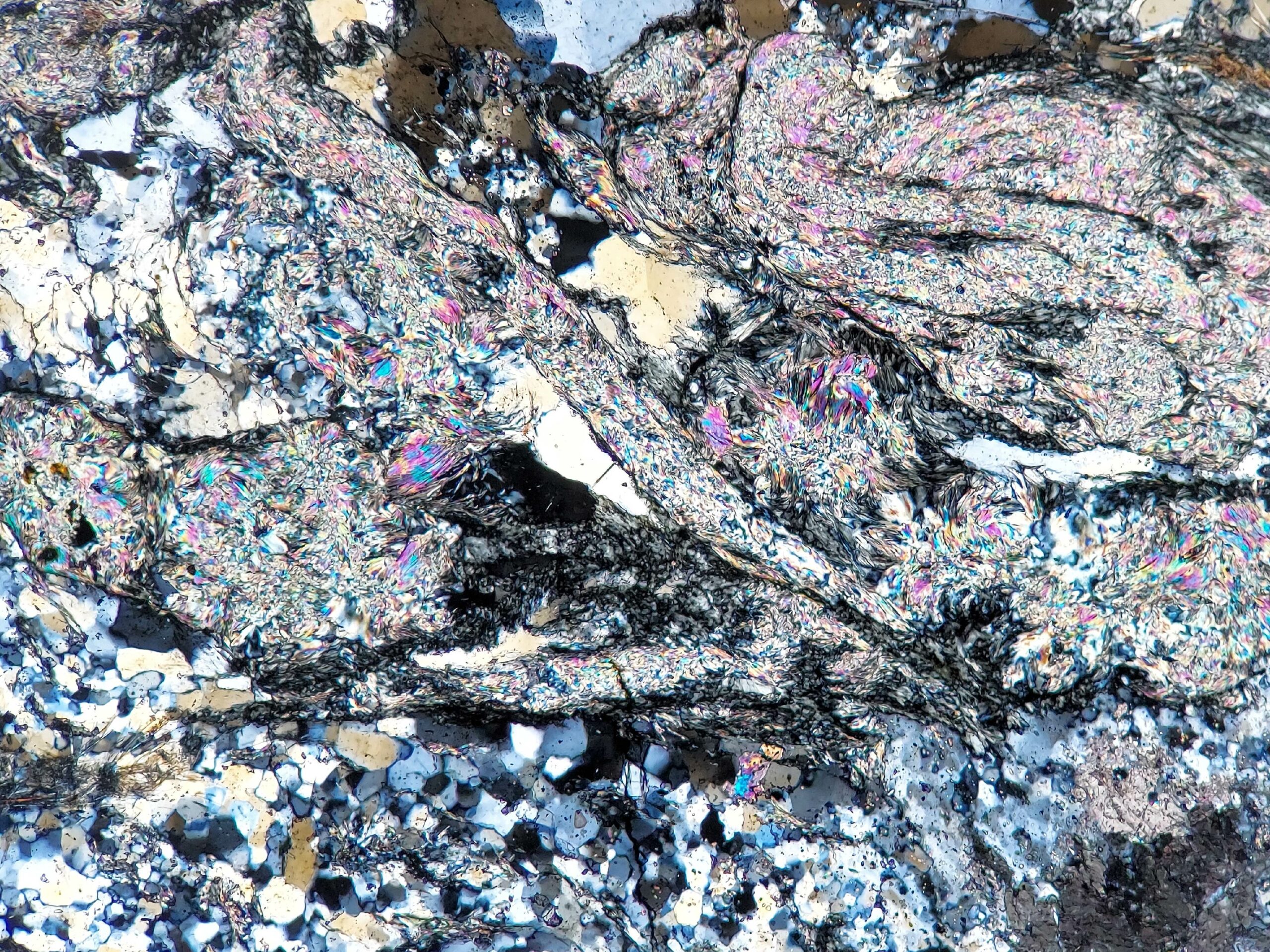
Fig 3.1.8 Serpentinite
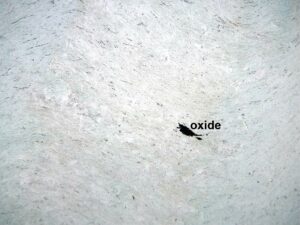
In PP light, a casual look at this thin section of a serpentinite from coastal California might appear to show a single black oxide grain – is there anything else present? But XP reveals the beauty of this rock, with its folded bands of light- to dark-gray serpentine. Reexamine it in PP, and closer inspection reveals serpentine’s slight variations in shading, from colorless to extremely pale-green or tan. FOV = 3.5 mm.
|


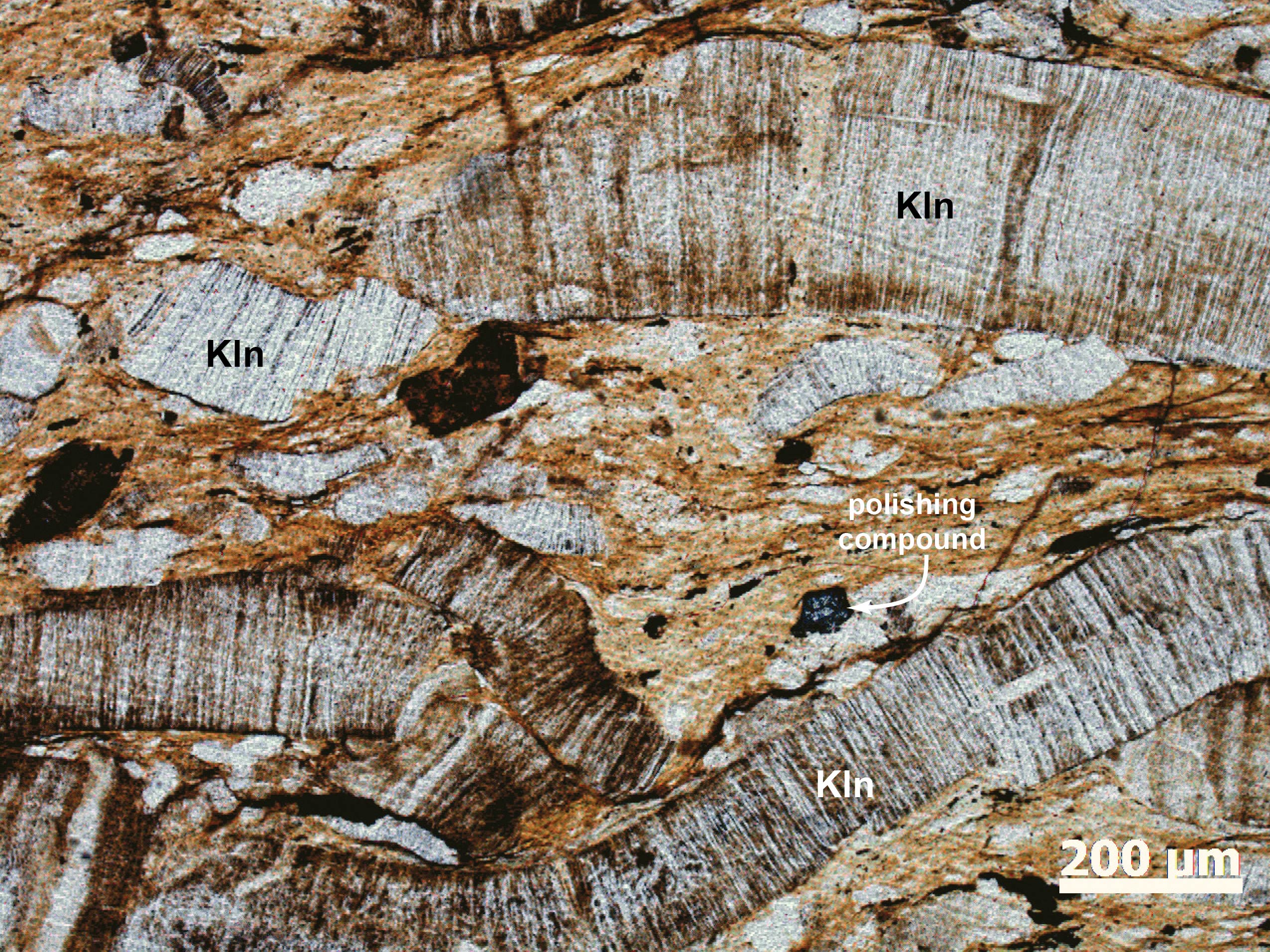
3.2 Clay Minerals
Clays have highly variable compositions; most are hydrated aluminosilicates
Occurrence—Clay minerals form as products of weathering and are abundant in a variety of sedimentary rocks (Figs 3.2.1, 3.2.2, 3.2.3, 3.2.4) and in soils. They also form in secondary veins, and as fine grained alteration products on or in coarser grained minerals (Fig 3.2.6).
In thin sections clay minerals may appear as replacement patches in feldspars, micas, and in many other silicates. Incipient alteration to clays can turn normally clear minerals cloudy (Fig 3.2.5). Generally, clay minerals are too fine-grained to see individual grains.
Distinguishing Features—In sedimentary rocks, clays appear as fine clasts, with up to 2nd-order interference colors. In altered rocks, clays normally appear as aggregates of fine scales or shards, or just discoloration in or on other minerals. Clays are normally colorless in thin section but fine grain size may give them a darker gray or brown hue.
Similar Minerals—All the many different clay minerals are similar, and many other secondary minerals, such as zeolites, may be mistaken for clays. Glauconite, sometimes considered a clay mineral, but more appropriately grouped with micas, is a conspicuous green mineral found in some sandstones.
Optical Properties—Most clay minerals are monoclinic; biaxial (- or +), although a few are triclinic, orthorhombic, or even hexagonal (uniaxial). Indices of refraction are normally 1.5-1.6, so relief is low. Some coarse clays may display up to middle 2nd-order interference colors, but kaolinite has very low birefringence. Fine-grained clay masses may appear nearly isotropic. In most cases, grain size is to small to permit interference figures.


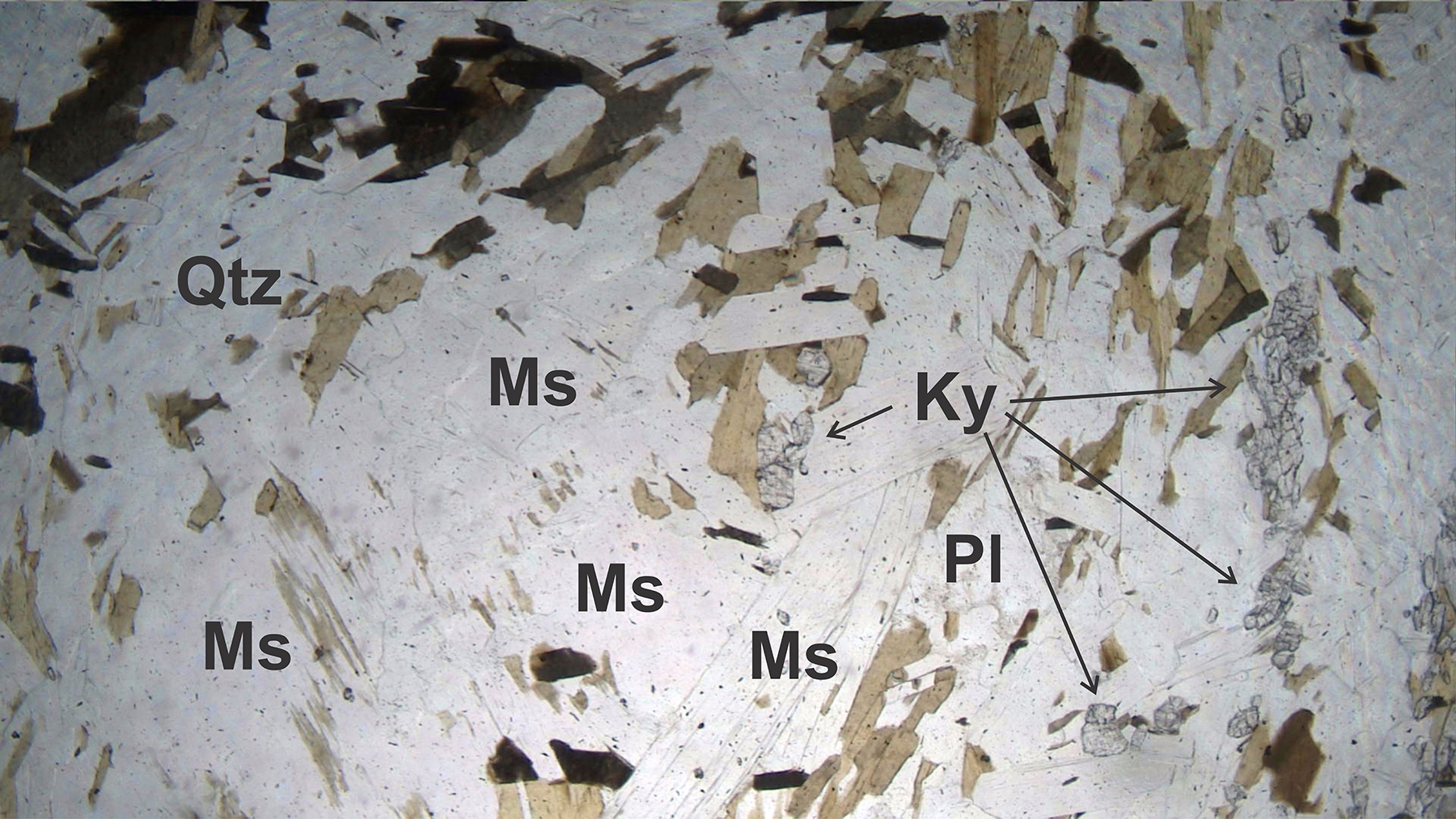
3.3 Pyrophyllite
Al2Si4O10(OH)2
Occurrence—Pyrophyllite is found in low- and medium-grade metamorphosed shales; more rarely as a secondary mineral in felsic volcanics and as a vein or ore mineral in metabauxites. It is typically fine grained and difficult to identify in thin sections of common rocks.
Distinguishing Features—Pyrophyllite looks much like muscovite. It is colorless in thin section, has one perfect cleavage, and has up to 3rd-order interference colors.
Patches or aggregates with curved, radial, or distorted crystals are common. Fine-grained aggregates are also possible. Twinning may be present but is difficult to see. In some cases, pyrophyllite displays bird’s-eye extinction (link to video).
Similar Minerals—Muscovite and talc may be confused with pyrophyllite, but both muscovite and talc have a smaller 2V, and muscovite crystals are normally not curved or distorted. Pyrophyllite is always associated with quartz. Talc is never found in aluminous rocks. Kaolinite has lower birefringence.
Optical Properties
■■ Triclinic; biaxial (-)
■■ 2V = 53° to 62°
■■ α = l.554, β = 1.588, γ= 1.601
■■ δ = 0.045-0.048. High birefringence yields up to 3rd-order interference colors; sections parallel to cleavage show white or gray colors.
■■ Cleavage flakes show a centered biaxial figure. Relief is low to moderate. Extinction is parallel (or nearly so) to cleavage and long dimension
■■ Slow direction is parallel to good cleavage.
|
 |
|
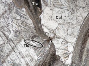
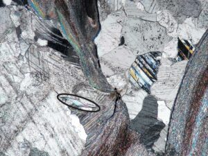
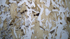
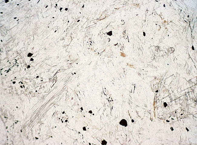
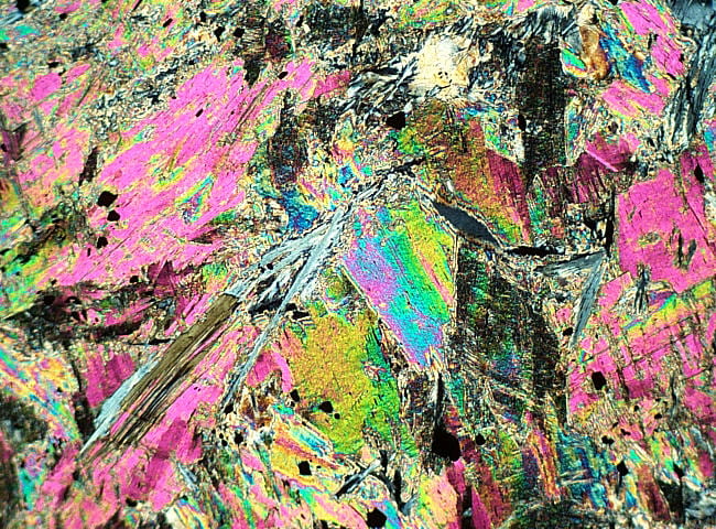
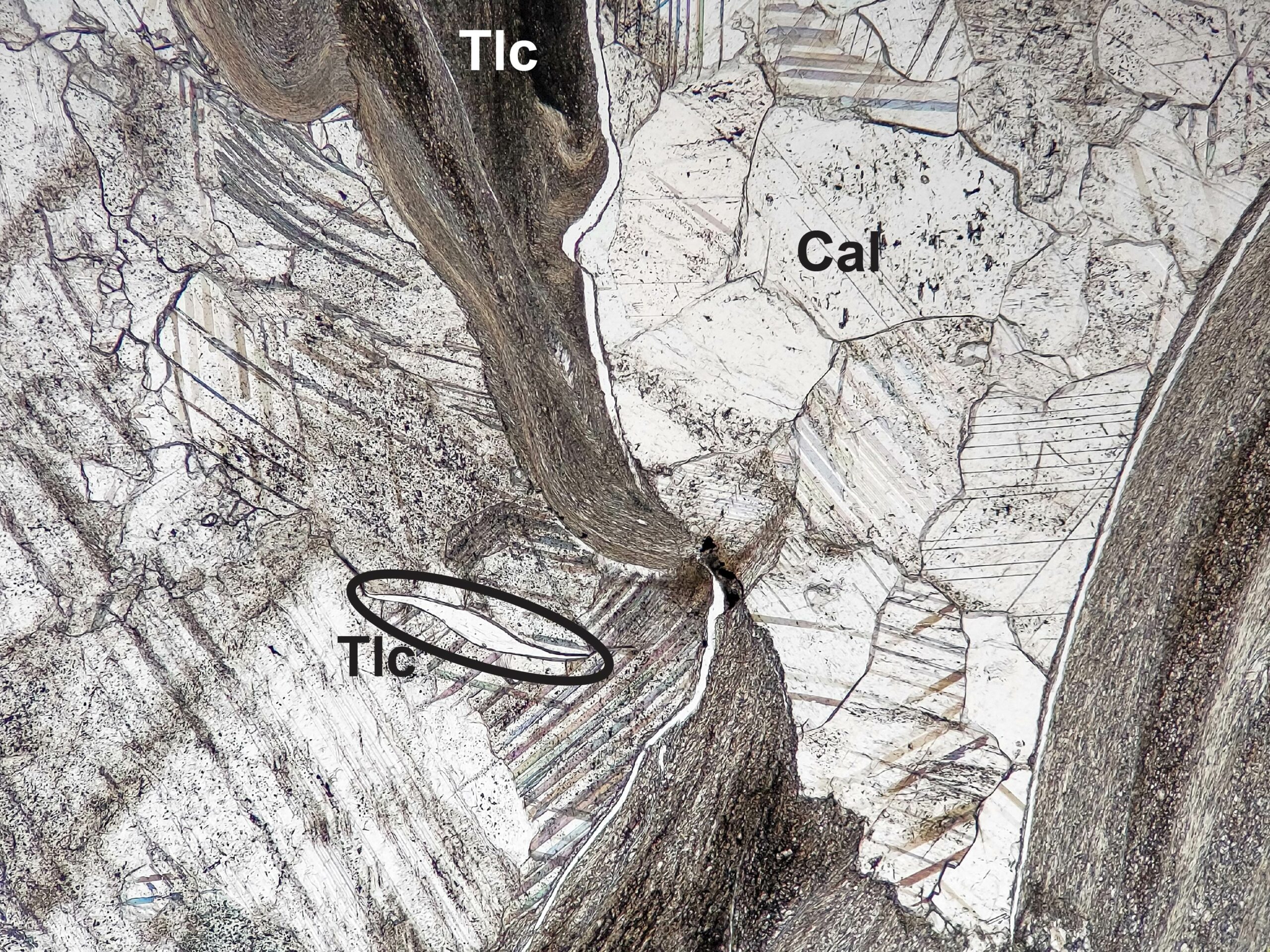
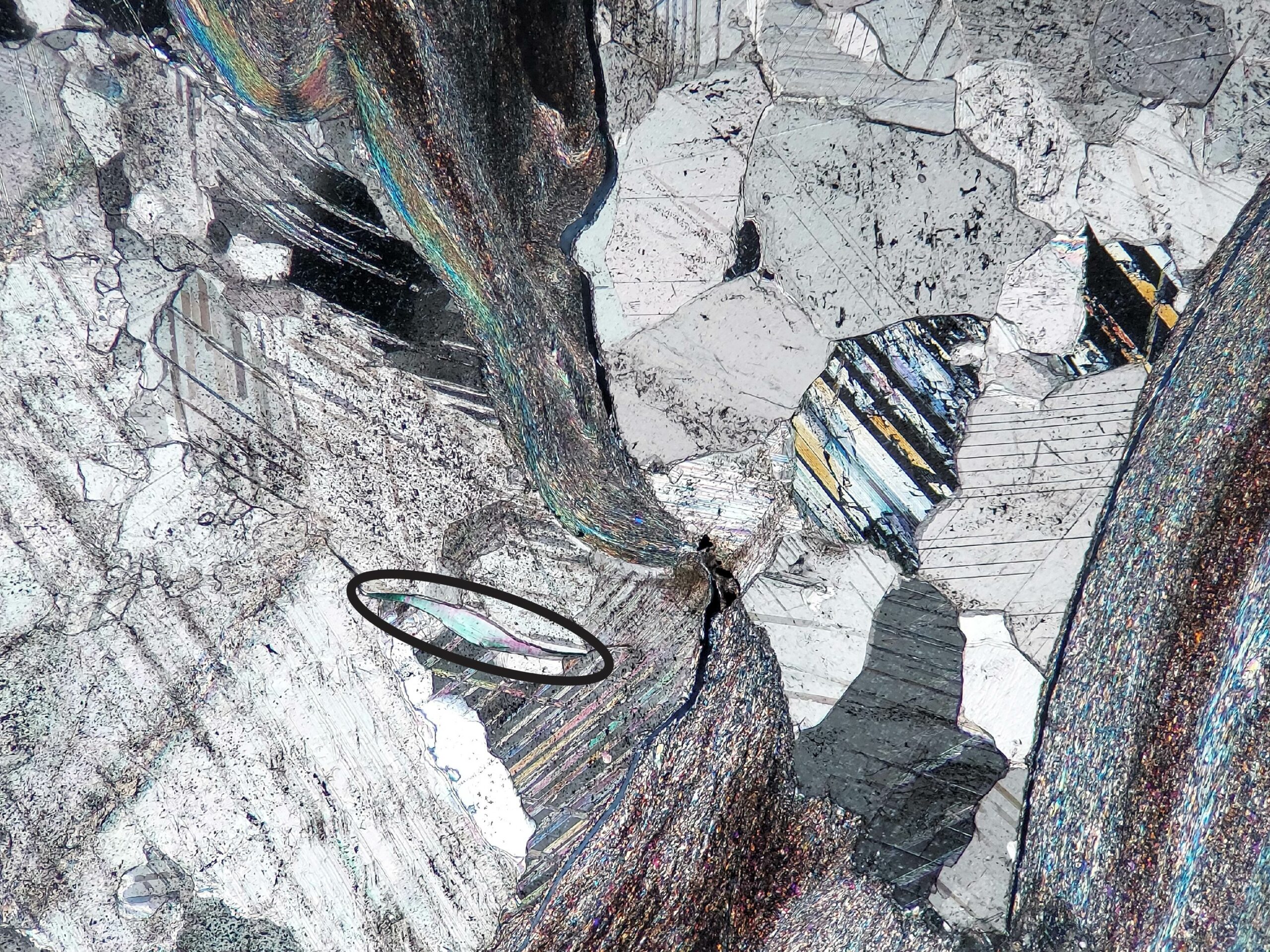
3.4 Talc
Mg3Si4O10(OH)2
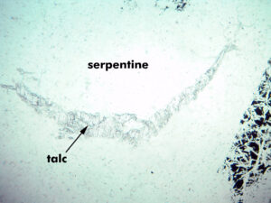
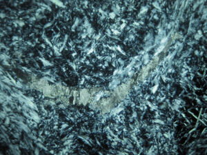
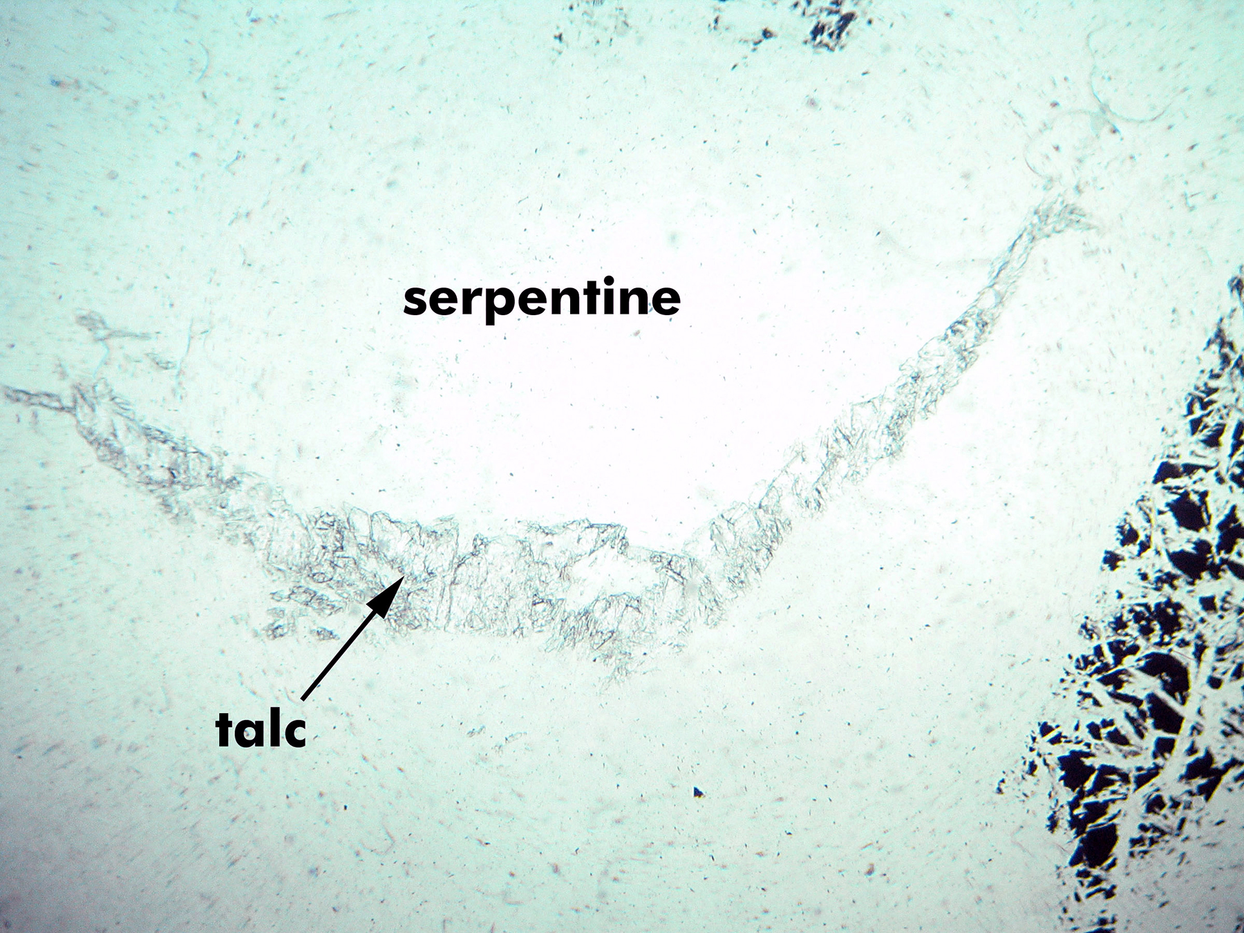
 Occurrence—Talc is a major mineral in some low-grade metamorphic rocks, including marbles and ultramafic rocks. It is a secondary mineral in altered mafic and ultramafic igneous rocks where it may be found with serpentine.
Distinguishing Features—Talc is a colorless sheet silicate that displays 3rd-order pastel and patchy interference colors. The pastel colors and rock associations are keys to identification.
Talc typically forms platy masses or fine to coarse fibrous aggregates with subparallel alignment. Bent fibers and shreds are common.
Talc has one good cleavage.
Similar Minerals—Talc resembles muscovite and pyrophyllite, but has higher-order interference color, lower 2V, and does not occur in aluminous rocks that are likely to have muscovite or pyrophyllite. Pyrophyllite is associated with quartz, whereas talc-quartz assemblages are rare. Brucite is uniaxial (+). Gibbsite is biaxial (+), has lower birefringence, and has oblique extinction.
Optical Properties
■■ Monoclinic; biaxial (-)
■■ 2V = 0° to 30°
■■ α = 1.539-1.550, β = 1.589-1.594, γ = 1.589-1.596
■■ δ = 0.046-0.050. Maximum interference colors are upper 3rd-order
■■ Cleavage flakes typically show low-order interference colors. Relief is low.
■■ Extinction angle is commonly just a few degrees, so extinction is nearly parallel to cleavage
■■ Length slow
|
|
 |
|
|
|
|
|
|

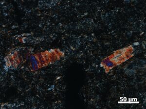
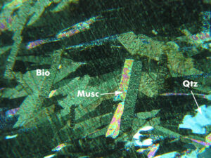
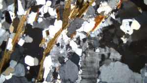
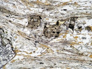
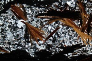
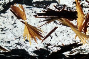
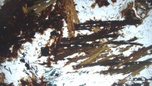
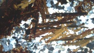
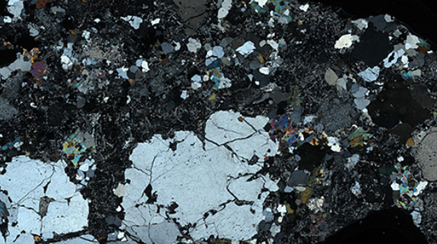
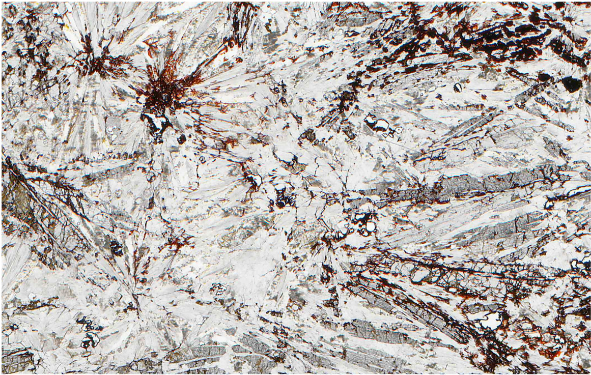
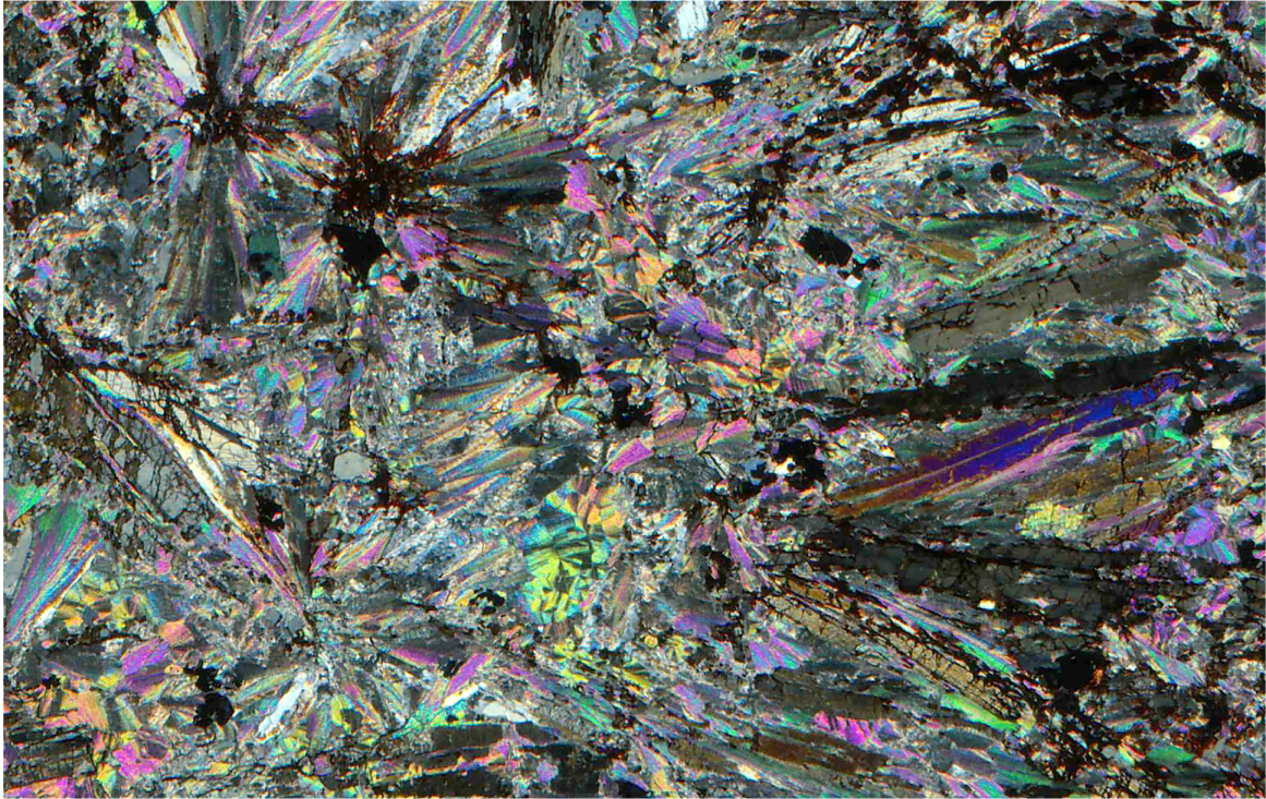
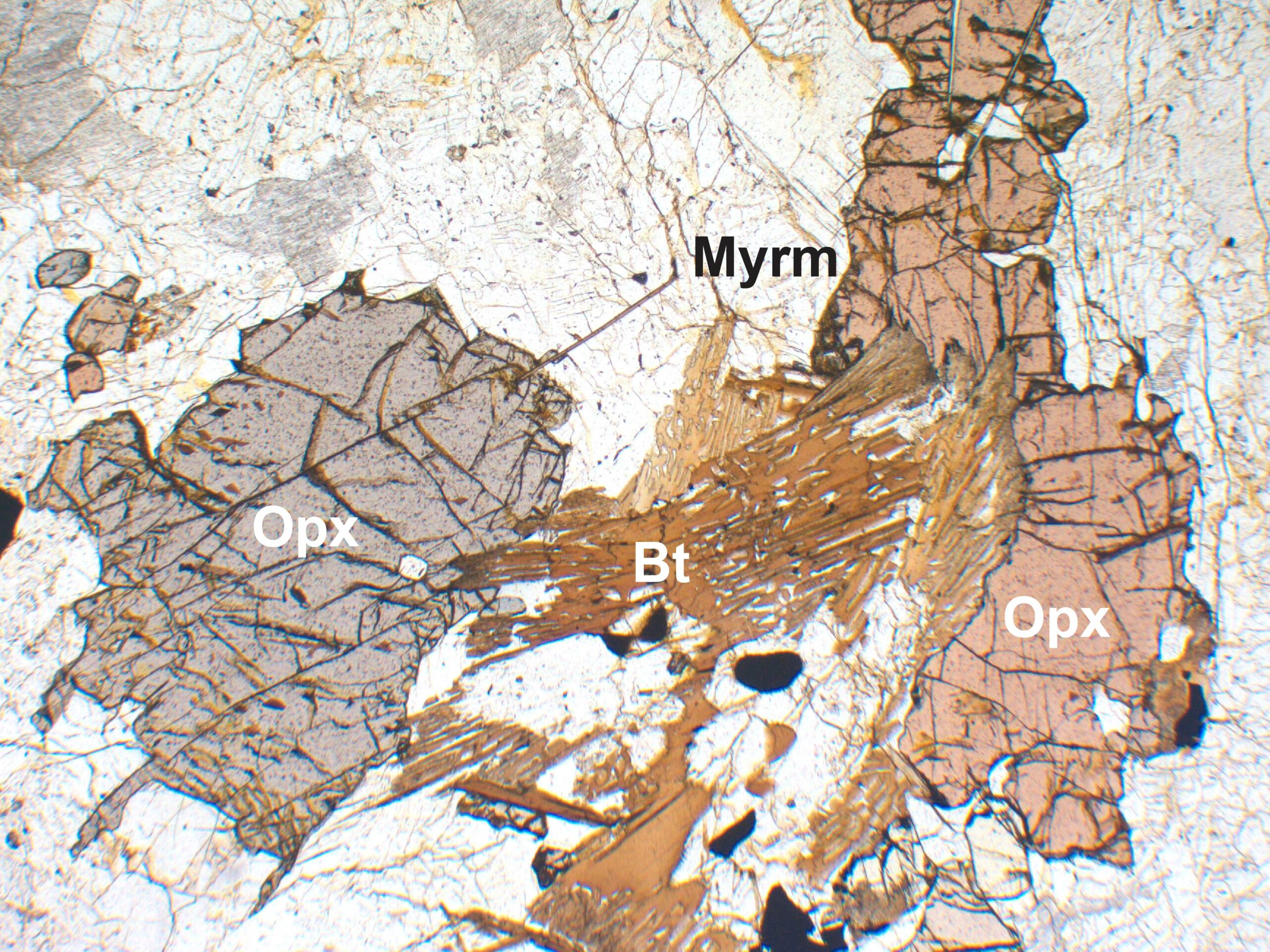
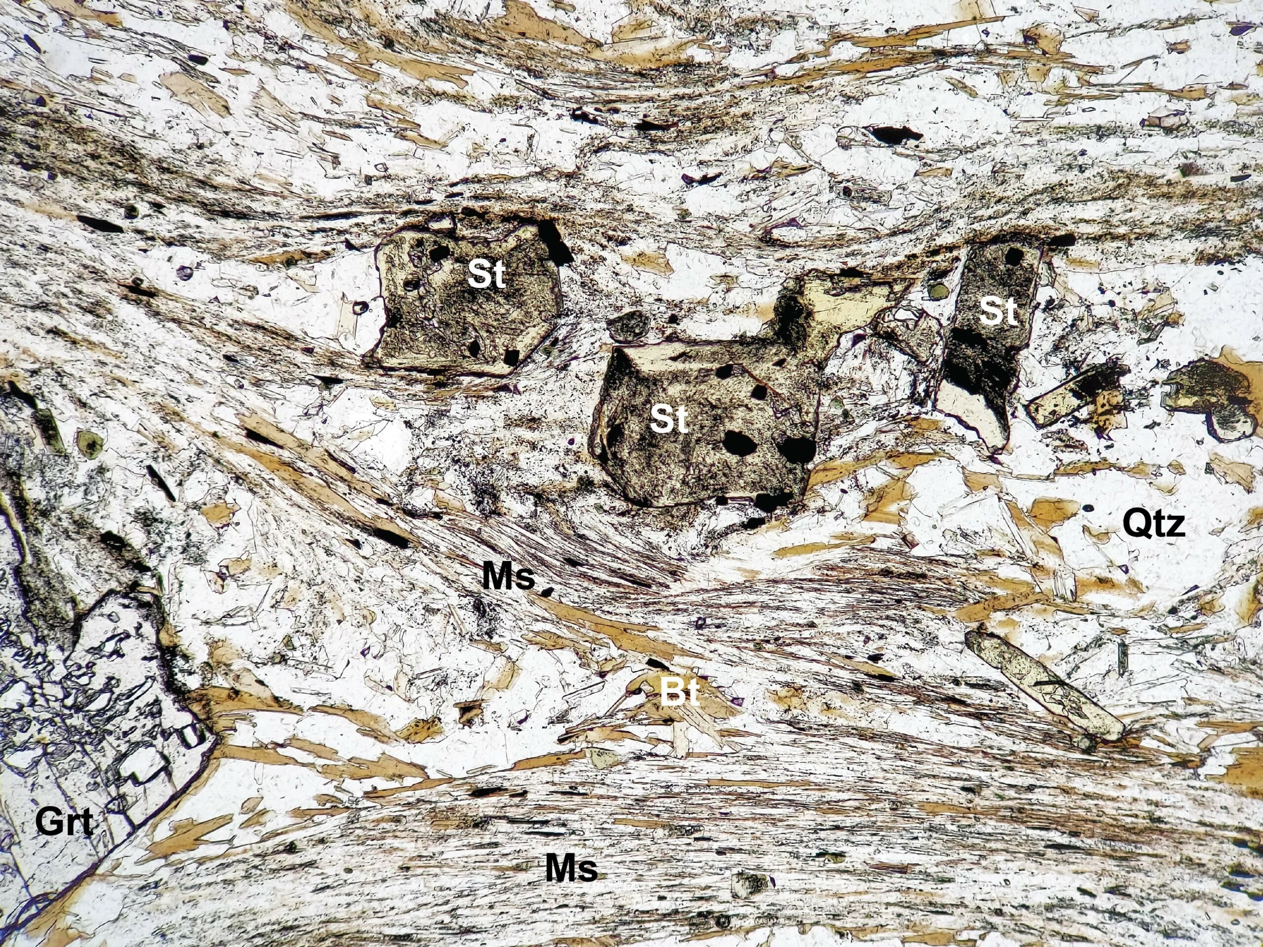
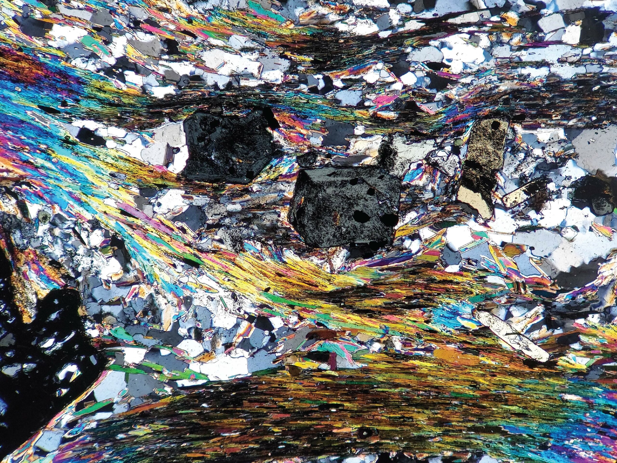
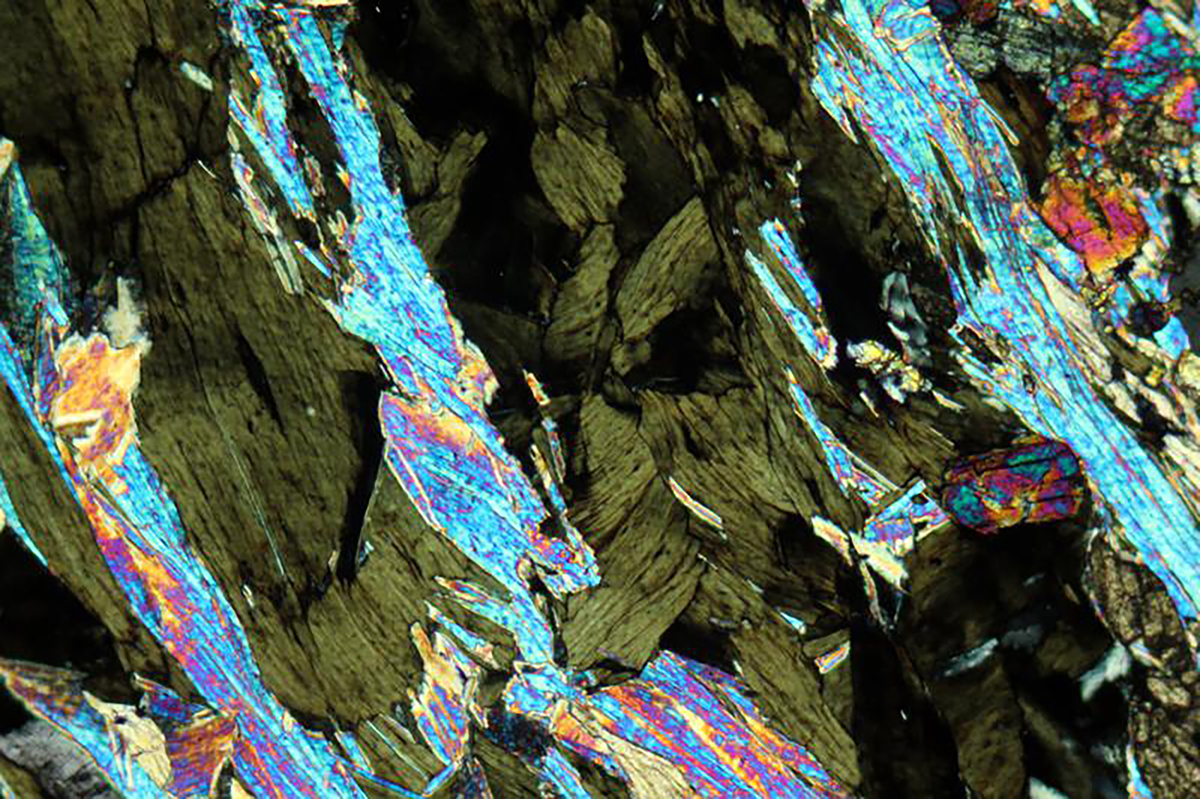
3.5 Biotite
K(Mg,Fe)3AlSi3O10(F,OH)2

Occurrence—Biotite occurs in a wide variety of igneous and metamorphic rocks; it is quite rare in sedimentary rocks, but appears occasionally in immature sedimentary rocks.
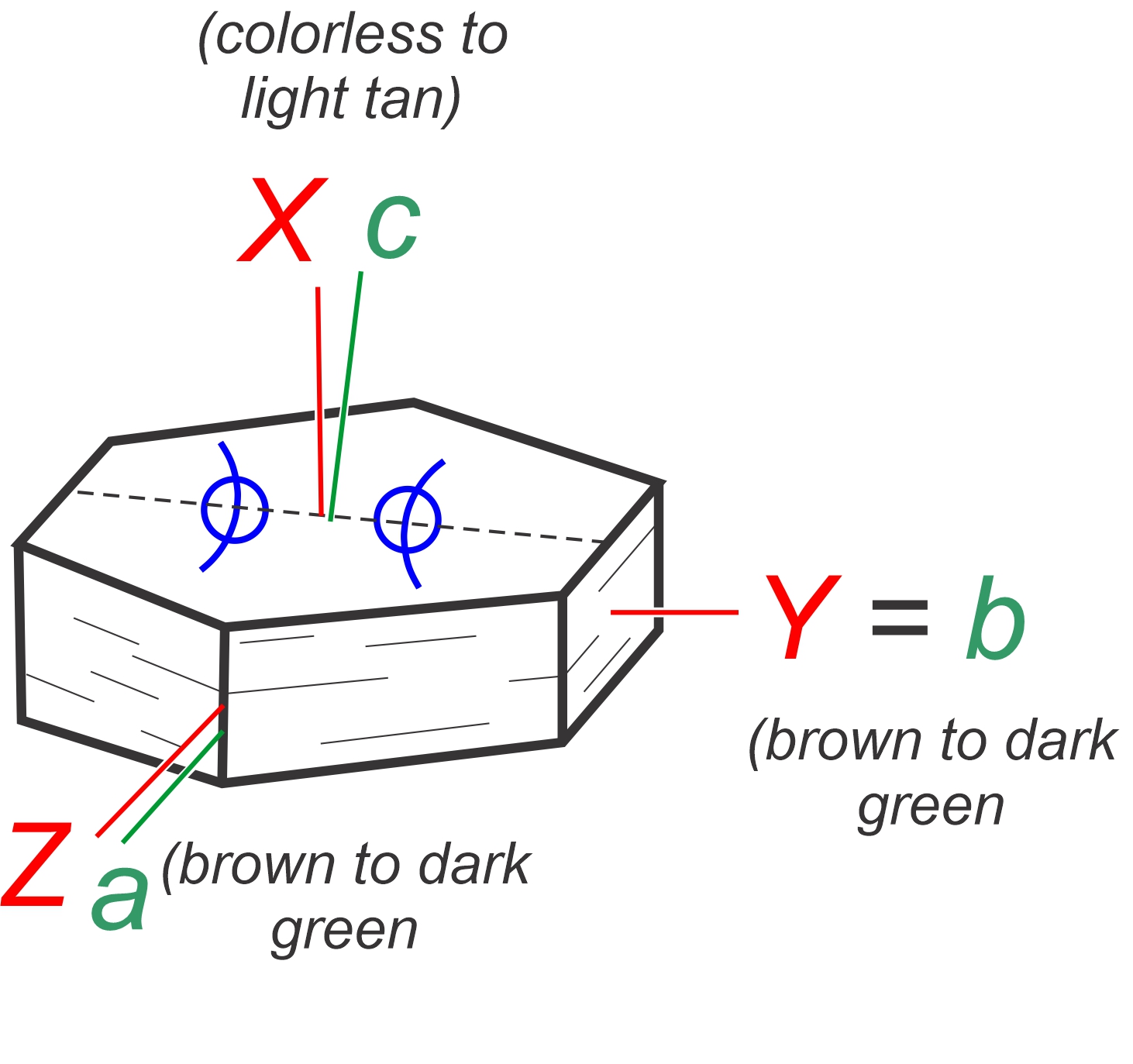
Distinguishing Features—Biotite is typically brown to yellow in thin section, but red or green hues are not uncommon. It is pleochroic and shows one excellent cleavage.
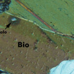
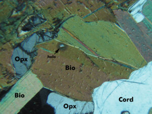
Biotite grains have mottled, or bird’s-eye, extinction in many/most thin sections (link to video). They may contain pleochroic halos around small included crystals of zircon or monazite.
In thin section, biotite commonly occurs as elongate rectangular flakes with a single cleavage. More equant plates or tabs are common too, depending on grain orientation. Rarely, flakes show hexagonal symmetry. Flakes commonly combine to form masses or aggregates, and can be bent. Twins may be present but can be difficult to see.
Similar Minerals—Biotite is distinguished from hornblende by cleavage (one cleavage, not two), color (usually more brown or red brown than green), relief (lower), and a smaller extinction angle. Amphiboles have two cleavages at about 60° and 120°, biotite has one cleavage. Biotite is difficult to distinguish from stilpnomelane, a brown brittle mica. Stilpnomelane occurs in low-temperature, high-pressure rocks where biotite is unstable, and gives a pseudouniaxial figure and has higher relief.
Color—Most biotites are pleochroic (X = colorless, light tan, pale greenish brown, or pale green, Y=Z= brown, olive brown, dark green, or dark red-brown). Phlogopite (Mg-biotite) is colorless to pale brown and occurs in Fe-poor rocks such as calc-silicate rocks.
Optical Properties
■■ Monoclinic (pseudohexagonal); biaxial (-)
■■ 2V = 0° to 33° for most biotite
■■ α = 1.571-1.616, β = 1.609-1.696, γ = 1.610-1.697; moderate relief
■■ δ = 0.028-0.081, strong interference colors range up to 2nd-order red, but they may be hard to see due to the color of the mineral
■■ Flakes lying on cleavage show very low-order colors, largely masked by the mineral’s color
■■ Interference figures may appear uniaxial depending on composition
■■ Normally parallel extinction, but extinction angle may be up to 3°
■■ Distorted plates show wavy extinction
■■ Cleavage direction is slow
|
 |
 |
|
|
|
|
|
|


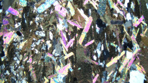
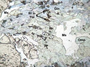
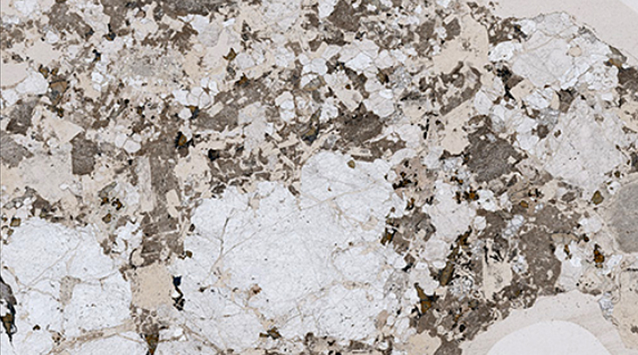
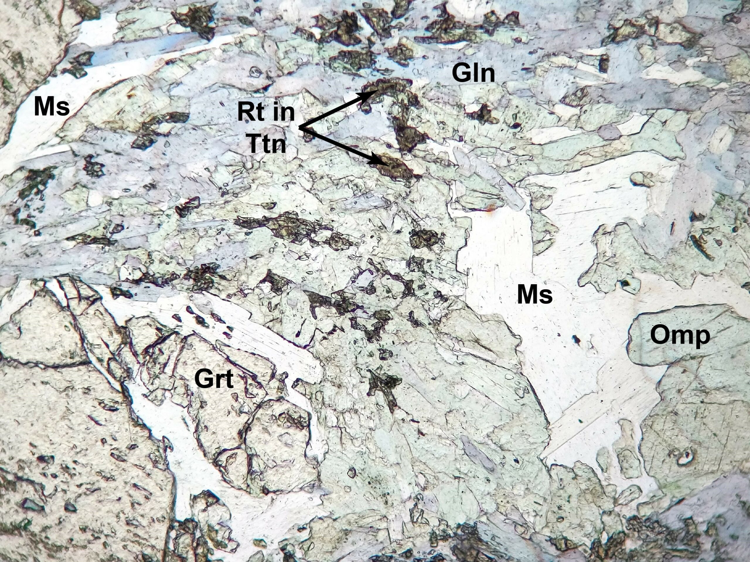
3.6 Muscovite
KAl2(AlSi3)O10(F,OH)2
 Occurrence—Muscovite is found in felsic to intermediate igneous rocks, in a wide variety of metamorphic rocks and, less commonly, in immature sedimentary rocks. A fine grained variety of muscovite (sericite) forms as an alteration product of feldspars and a few other minerals. High pressure muscovite is called phengite.
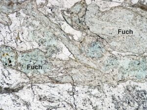
Distinguishing Features—Muscovite is almost always colorless mica with moderate relief. A rare Cr-bearing variety of muscovite, called fuchsite, has a diagnostic green color.
Muscovite forms tabular crystals, rectangular flakes, or laths when coarse; scaly aggregates or shreds when fine. It generally shows one excellent cleavage. Twinning is common but hard to detect.
Muscovite has 2nd-order interference colors, up to yellow or red. Under crossed polars it commonly displays bird’s-eye extinction (link to video).
Similar Minerals—Some other rarer white micas (paragonite and margarite) and lepidolite have similar properties and cannot be distinguished in thin section. Chemical analyses, XRD, or other tests are needed to distinguish them. Some clay minerals, too, appear superficially like muscovite in thin section.
Optical Properties
■■ Monoclinic; biaxial (-)
■■ 2V = 35° to 50°
■■ α = 1.552-1.770, β = 1.582-1.619, γ = 1.588-1.624; moderate relief may vary with stage rotation
■■ δ = 0.036-0.054; up to 2nd-order yellow or red interference colors; sections parallel to cleavage show 1st-order white
■■ Cleavage fragments yield a nearly centered Bxa figures.
■■ Generally parallel extinction or a very small extinction angle
■■ Cleavage direction is length slow
|
 |
 |
|
|
|
|
|
|
|
|
|


3.7 Stilpnomelane
K(Fe2+,Mg,Fe3+)8(Si,Al)12(O,OH)27·n(H2O)
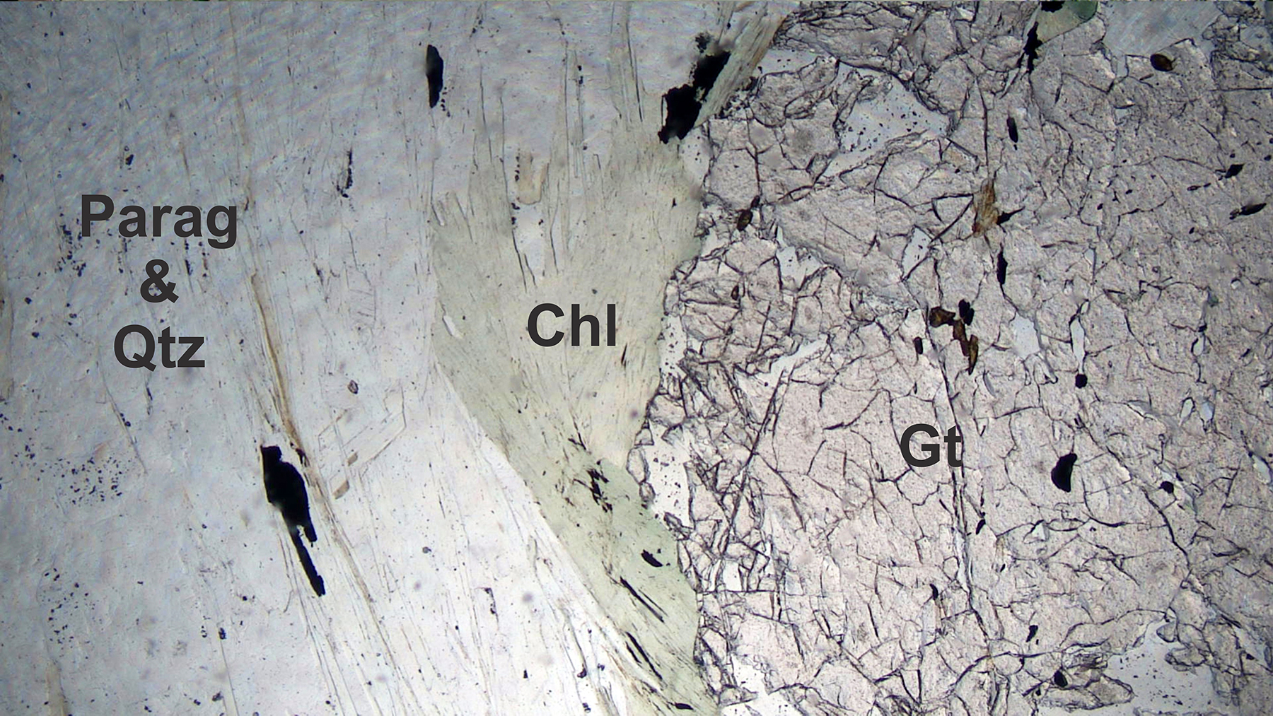
Occurrence—Stilpnomelane is common in low-grade high pressure metamorphic rocks. It is generally associated with chlorite, muscovite, garnet, actinolite, calcite, or epidote.
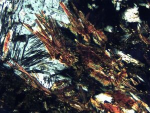
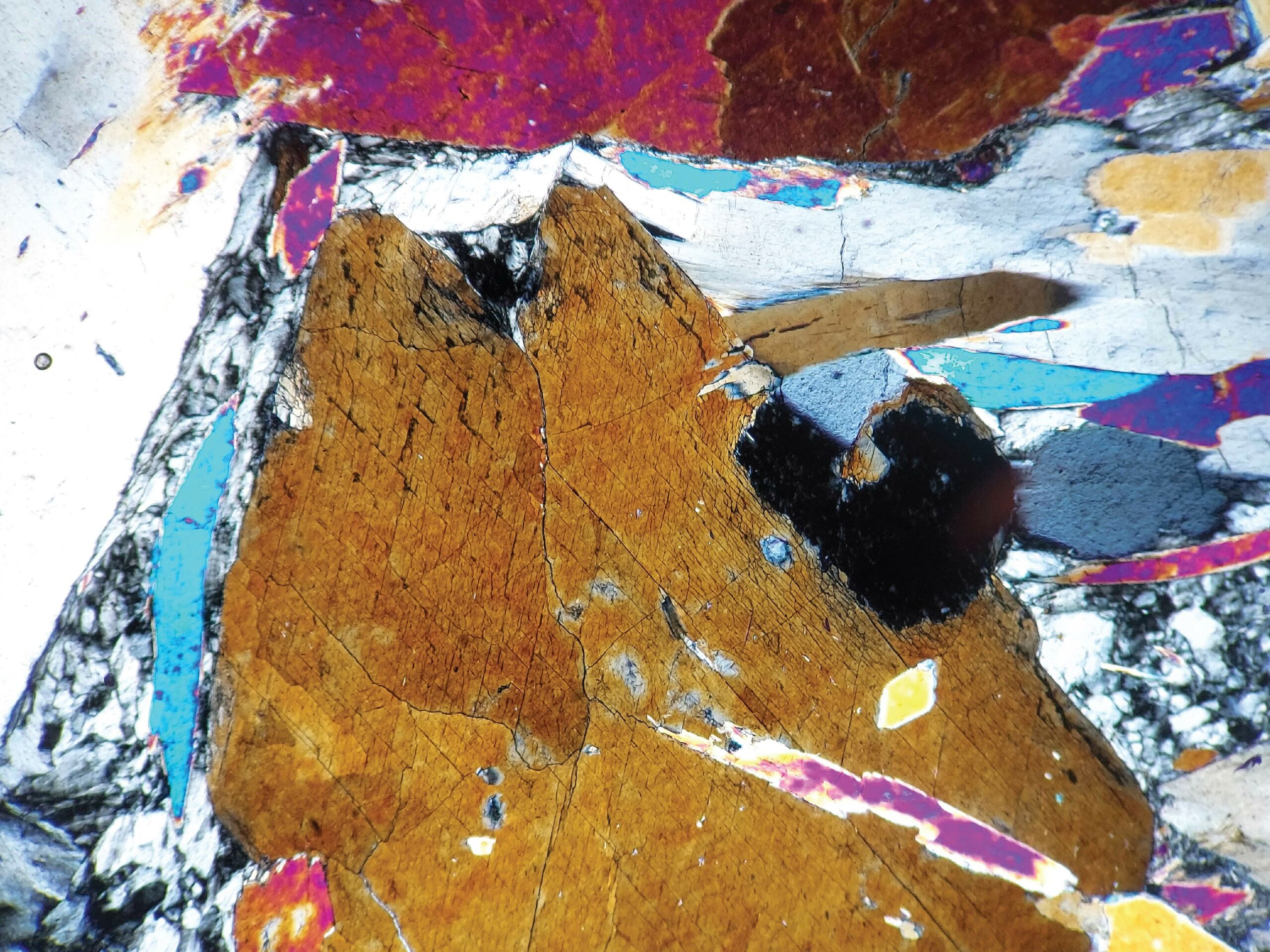
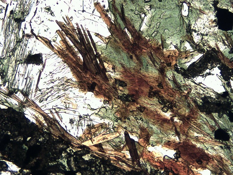
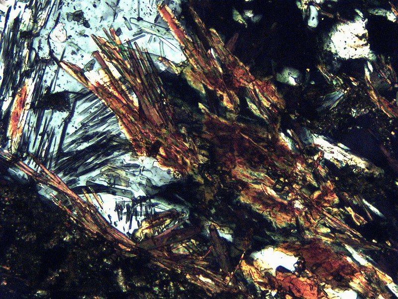
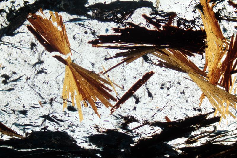
Distinguishing Features—Stilpnomelane is a high-relief dark brown, pale yellow, or green mica-like mineral with strong pleochroism. Its strong brown color and radiating habit are distinctive.
Stilpnomelane may form long elongate grains that in some cases seem to pile up on top of each other. It also occurs in sheaves or radiating aggregates/splays.
Stilpnomelane commonly shows two cleavages. It has one good and one fair cleavage that intersect at 90° in properly oriented crystals.
Similar Minerals—Stilpnomelane is hard to distinguish from biotite, but stilpnomelane generally has a deeper brown color. Stilpnomelane’s basal cleavage is less perfect than biotite’s and is intersected by an additional cleavage. Bird’s-eye extinction is absent or weakly present, and stilpnomelane’s birefringence may be greater than that of biotite.
Optical Properties
■■ Monoclinic; biaxial (-)
■■ 2V≈0; biaxial interference figure appears uniaxial
■■ α = 1.543-1.634, β = 1.576-1.745, γ = 1.576-1.745, but varies with composition
■■ δ = 0.030-0.110; birefringence varies with composition
■■ Maximum interference colors may be up to 6th-order, but the strong mineral color tends to mask them
■■ Relief is high, and may vary with stage rotation
■■ Extinction angle with the principle cleavage is close to 0°
■■ Principal cleavage direction is slow
|
 |
 |
|
|
|

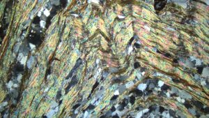
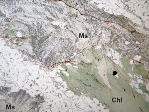
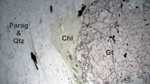
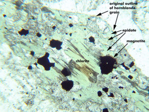
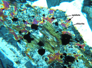
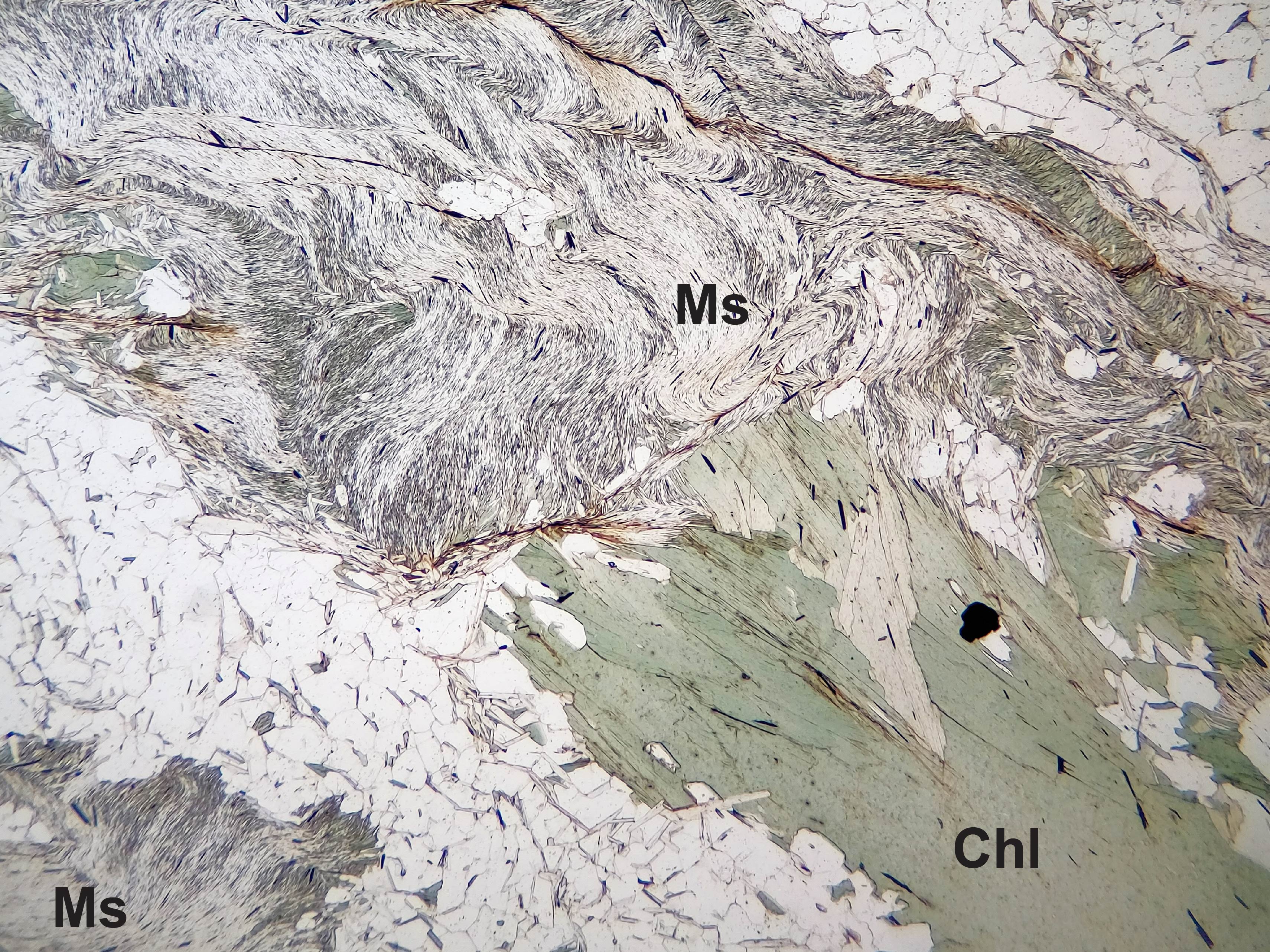
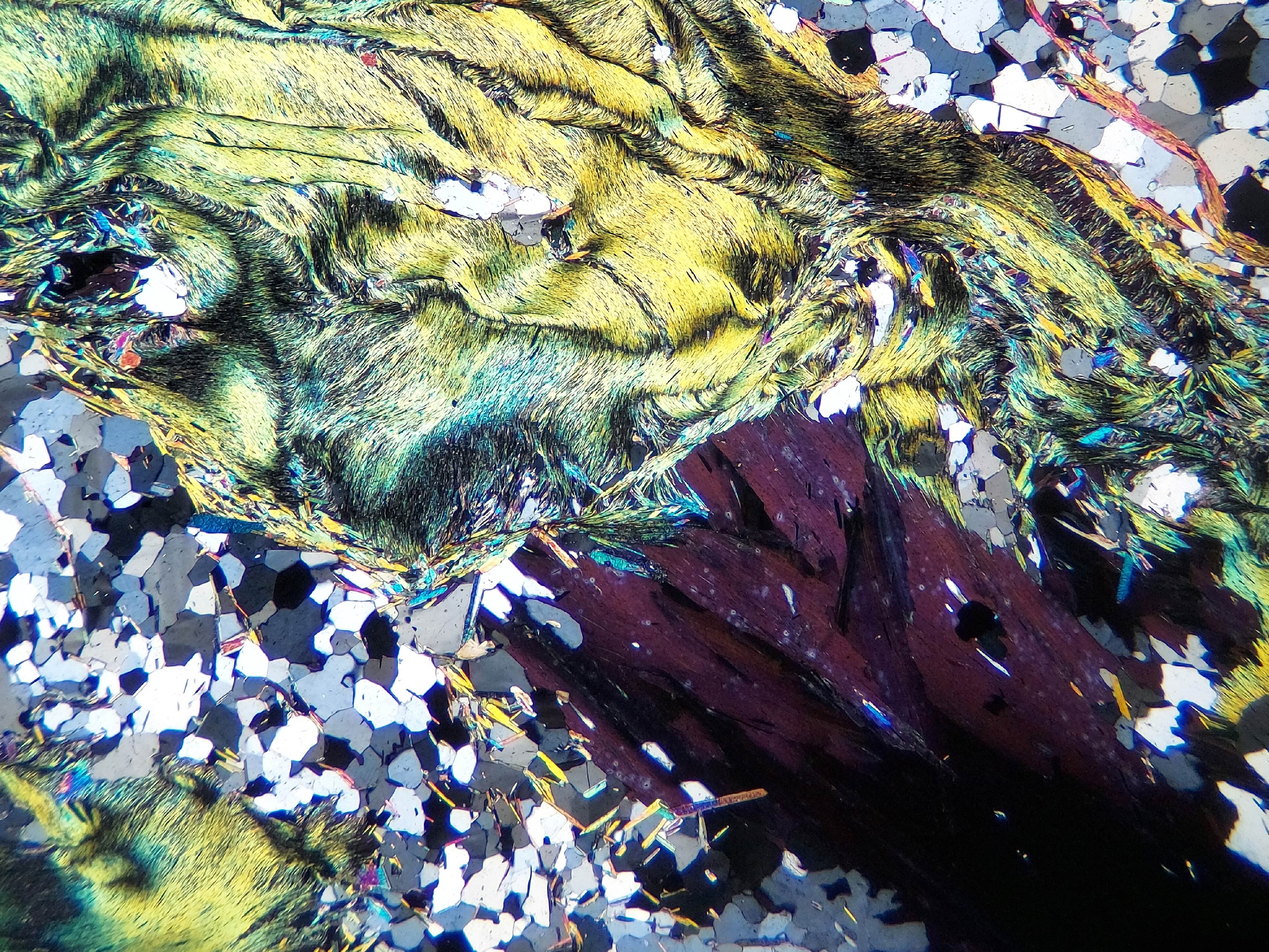
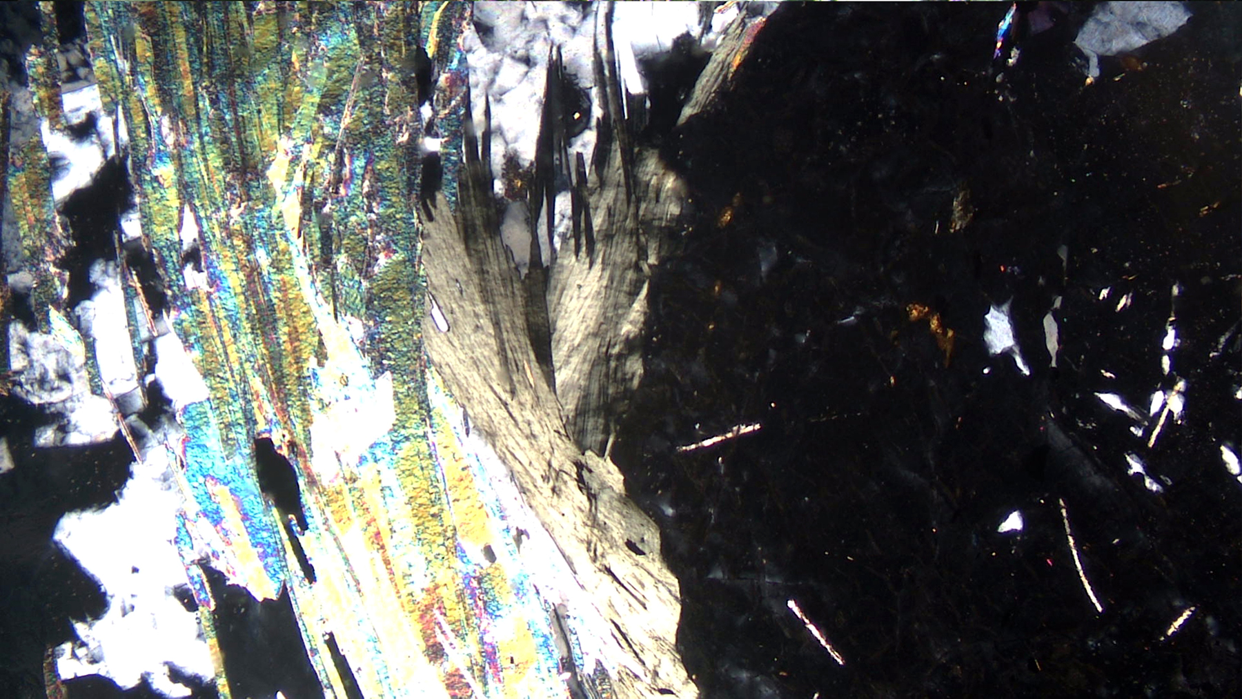
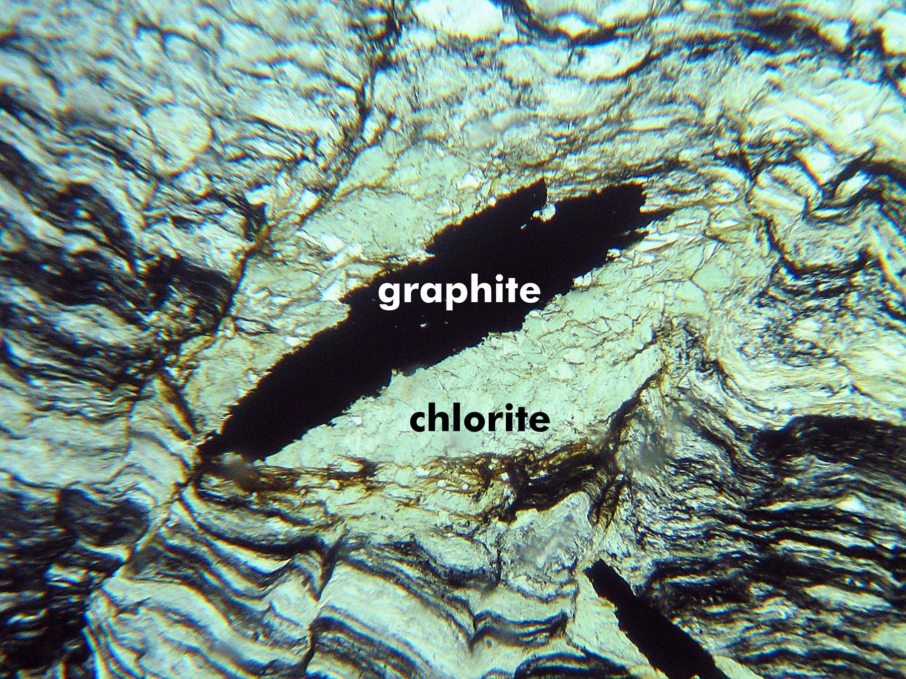
3.8 Chlorite
(Mg,Fe,Al)6(Si,Al)4O10(OH)8
 Occurrence—Chlorite is common in low- and medium-grade metamorphic rocks, both as a primary mineral and as a secondary mineral forming after biotite and other mafic silicates in both igneous and metamorphic rocks.
Distinguishing Features—There are many varieties of chlorite and properties vary somewhat.
In thin section, chlorite occurs as massive patches, flaky or scaly crystals, or thin to thick tabs. It is a common replacement for other minerals. In some cases it is pseudohexagonal. Polysynthetic twinning is common but generally hard to see.
Color is variable but normally light greenish or army green, more rarely shades of brown or yellow. Chlorite is normally pleochroic (although it may be faint) and displays low 1st-order or anomalous interference colors (blue, green, purple, or brown).
Chlorite has one excellent cleavage, but the cleavage may not be apparent in small grains.
Similar Minerals—Epidote, zoisite, pumpellyite and a few other minerals commonly exhibit anomalous interference colors, but are much higher relief, and generally don’t show green pleochroism. Chorite’s generally light-green color, form, and associations usually serve to distinguish it.
Optical Properties
■■ Monoclinic; biaxial (+) or (-)
■■ 2V = 0° to 50°
■■ α = 1.571-1.588, β = 1.571-1.588, γ = 1.576-1.597
■■ δ = 0.006-0.020; interference colors are very weak
■■ Anomalous interference colors (blue, purple, or brown, depending on Fe-Mg composition) are typical
■■ Rarely chlorite shows 1st-order light-yellow colors
■■ Relief is moderate to high
■■ Extinction angle varies from 0° to about 10°
■■ Crystals showing cleavage are length fast
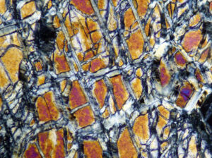
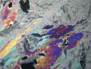
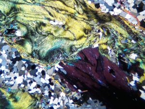
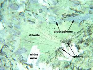
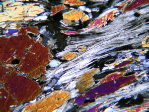
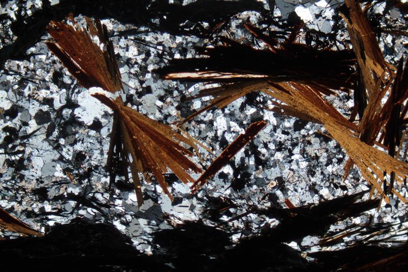
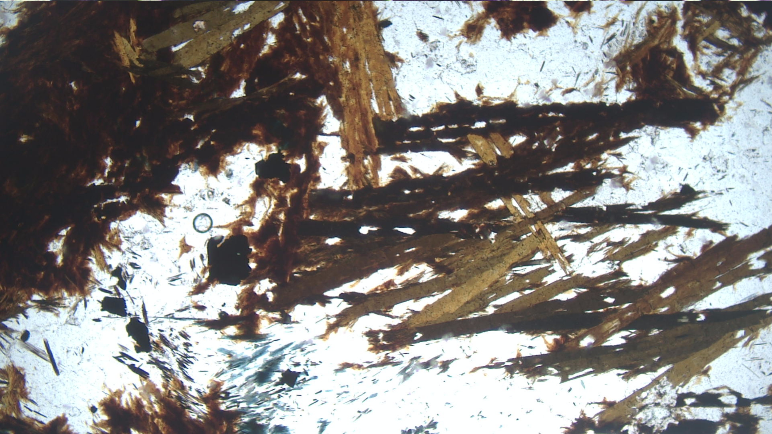
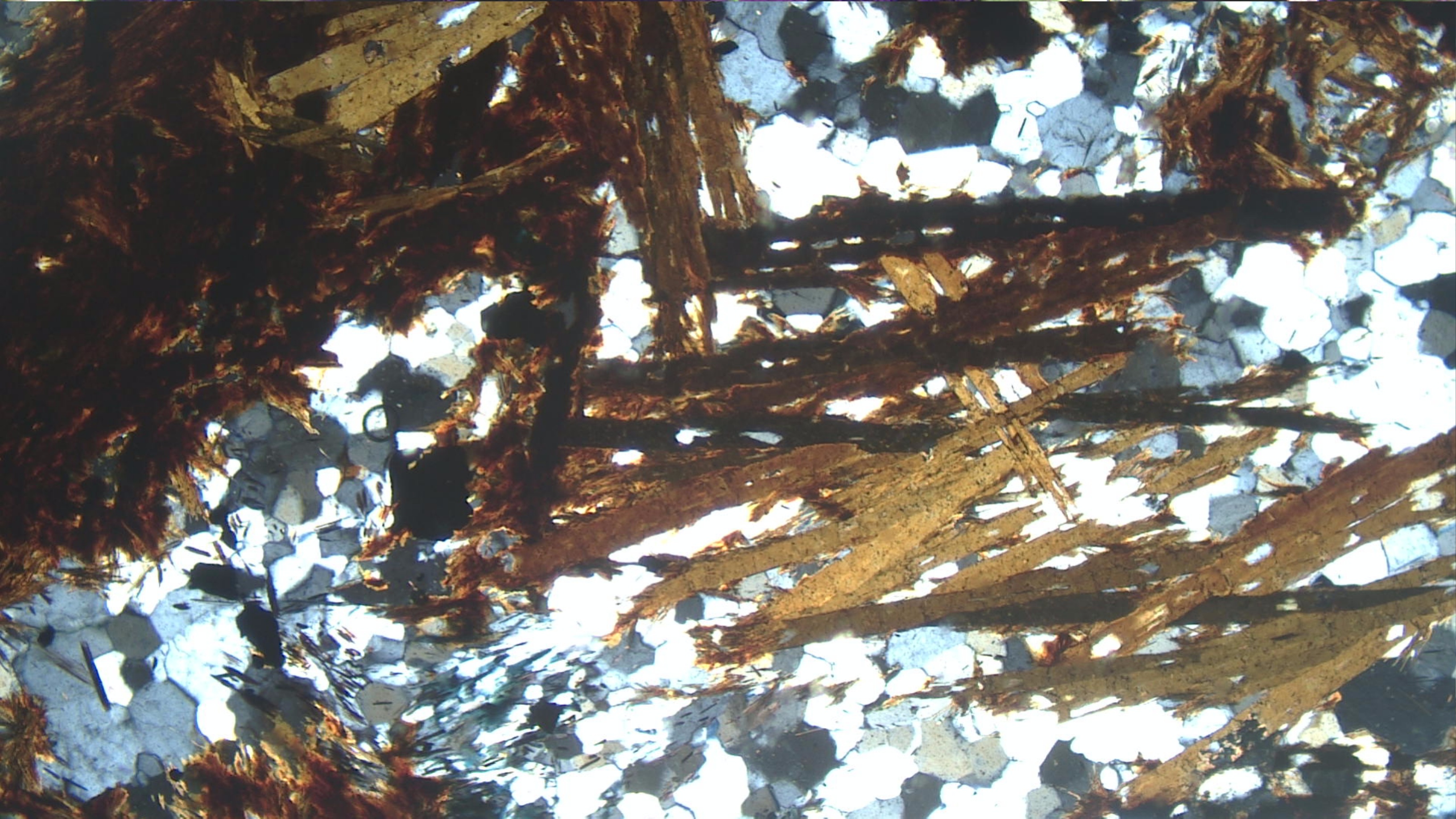
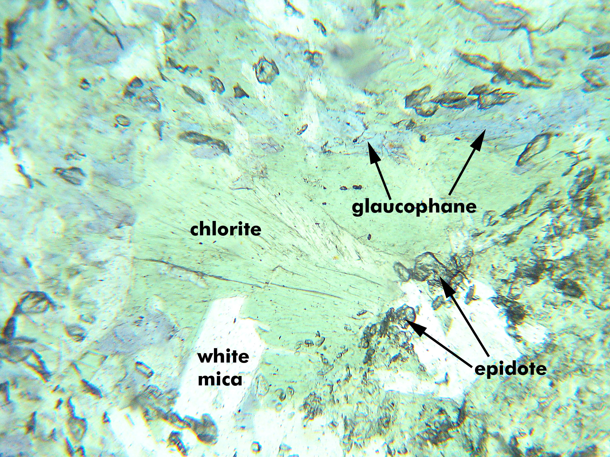
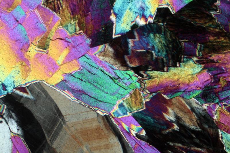
Fig 3.8.5 Blueschist
This blueschist from Panoche Pass, CA, contains pale green chlorite, blue glaucophane, colorless white mica flakes (bottom center and right) and many small high-relief epidote grains (PP). In XP light the chlorite shows anomalous interference colors, the glaucophane shows 2nd-order interference colors that are not generally typical of this mineral. The epidote grains are hard to pick out in XP but the mica flakes show typical mottled 2nd-order interference colors. FOV = 2.5 mm.
|
 |
 |
|
|
|
|
|
|
| Fig 3.8.5 Blueschist
This blueschist from Panoche Pass, CA, contains pale green chlorite, blue glaucophane, colorless white mica flakes (bottom center and right) and many small high-relief epidote grains (PP). In XP light the chlorite shows anomalous interference colors, the glaucophane shows 2nd-order interference colors that are not generally typical of this mineral. The epidote grains are hard to pick out in XP but the mica flakes show typical mottled 2nd-order interference colors. FOV = 2.5 mm. |
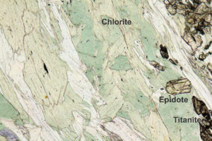
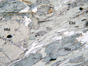
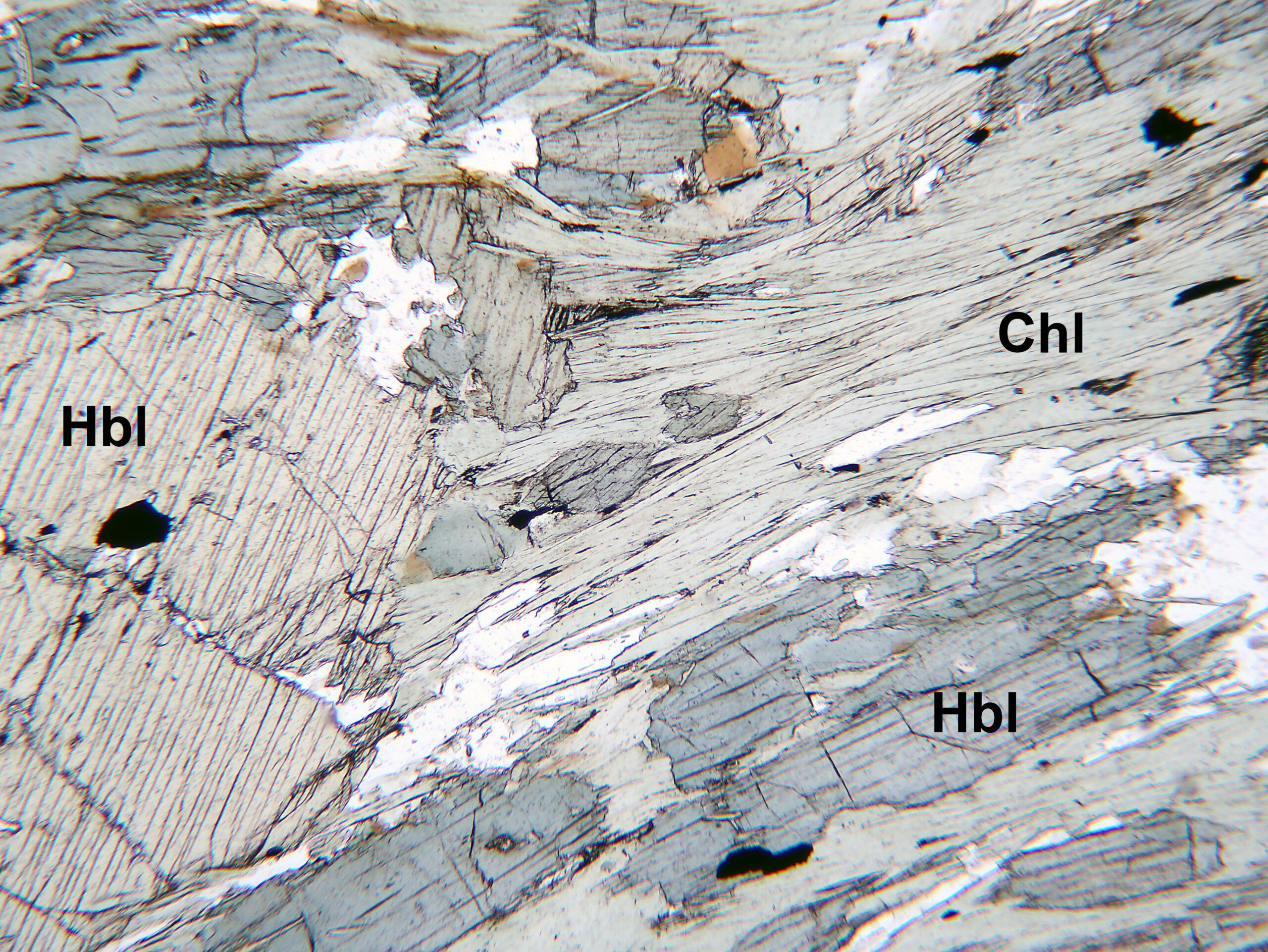
Fig 3.8.6 Amphibolite Chlorite (Chl) is pale green in PP and aligned in the foliation plane with blue-green hornblende (Hbl) laths. Colorless plagioclase and quartz are also visible along with a few grains of brown biotite. Chlorite displays anomalous 1st-order gray-blue colors in a distinctive texture in XP in contrast to the higher 1st-order red-orange to blue hornblende colors. The plagioclase is untwinned. This rock comes from near Hanover, New Hampshire. FOV = 2.5 mm.■■ Larger photos: PP XP |


3.9 Prehnite
Ca2Al2Si3O10(OH)2
Occurrence—Prehnite is a low-grade metamorphic mineral in mafic rocks (prehnite-pumpellyite facies) and also commonly occurs as an alteration product in basalts and other igneous rocks. It generally occurs in veins or vugs.
Distinguishing Features—Prehnite is colorless and has 2nd-order interference colors. It has one good cleavage. It commonly forms round globs or sheaf-like aggregates, but may show a “bow tie” or radiating structure. Fine polysynthetic twins in two directions might be seen in XP light.
Similar Minerals—Prehnite may be confused with lawsonite, but lawsonite has lower birefringence, higher relief, and different mineral associations (e.g., with glaucophane).
Optical Properties
■■ Orthorhombic; biaxial (+)
■■ 2V = 65° to 69°
■■ α = 1.611-1.630, β = 1.617-1.641, γ = 1.632-1.669
■■ δ = 0.021-0.039; maximum interference colors are low to upper 2nd-order Relief is moderate to high
■■ Extinction is parallel; may be wavy
|
 |
 |
