Ring silicates have structures in which all silicon tetrahedra are in hexagonal (6-membered) rings. Using this definition, tourmaline is the only common example. The very rare minerals dioptase, CuSiO2(OH)2, and benitoite, BaTiSi3O9, are also ring silicates.
Mineralogists sometimes group beryl and cordierite with tourmaline (as we have done in this book) because the two minerals, like tourmaline, contain hexagonal rings of tetrahedra. But, in beryl and cordierite, tetrahedral rings are connected by additional silicon tetrahedra that are not in rings.
6.1 Tourmaline
 Occurrence—Tourmaline is a common accessory mineral in granitic igneous rocks and in many metamorphic rocks, especially in mica schists. It is also found as a major mineral in some pegmatites and in clastic sedimentary rocks.
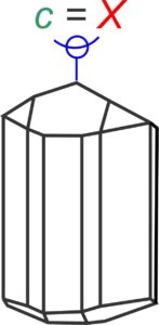
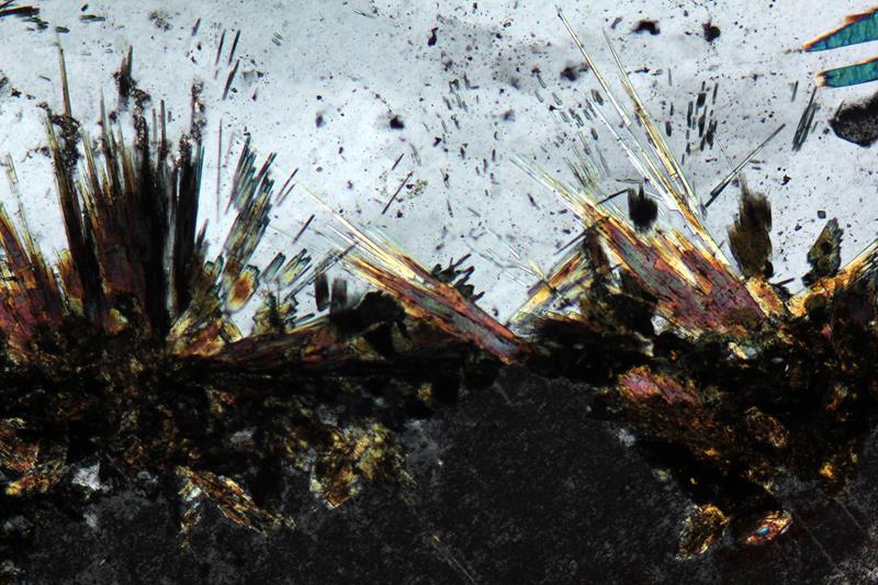
Distinguishing Features—Tourmaline generally occurs as small strongly colored grains with moderate- to high-relief. Stubby columnar or prismatic crystals with rounded triangular or nearly hexagonal cross sections are typical; columnar or fibrous radiating aggregates also occur. Tourmaline has no cleavage.
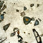
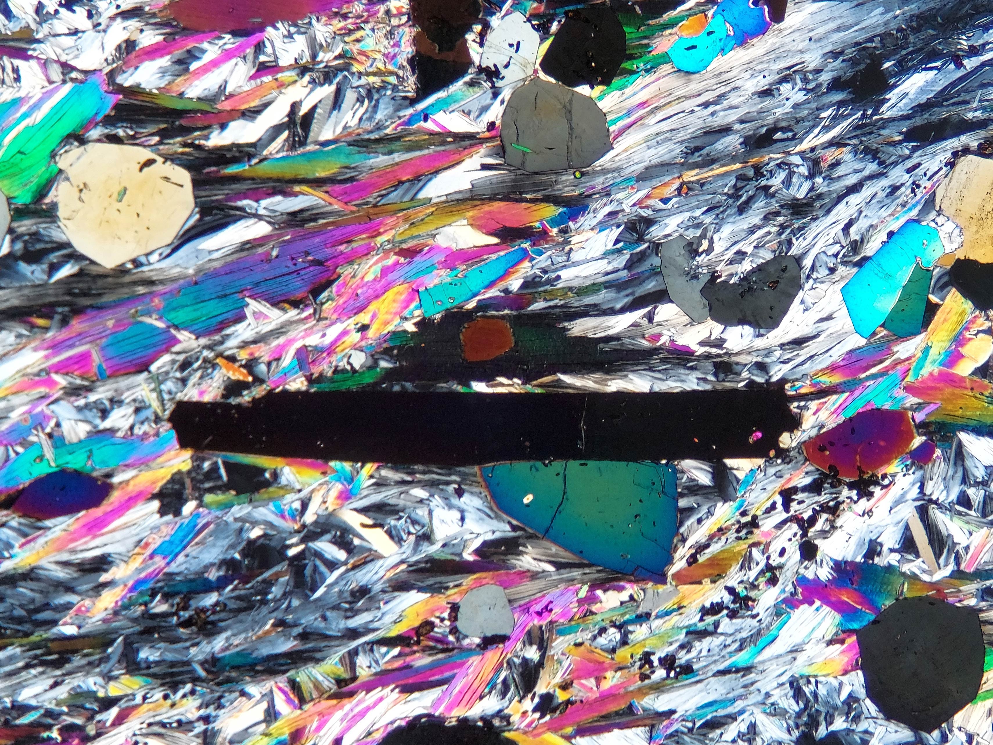
Color is variable; it may be brown, green, or blue, and less commonly yellow, red, pink, brown, or black. Tourmaline normally shows marked pleochroism and in many cases compositional zoning that causes color zoning. Elongated tourmaline crystals display their most intense colors when the long direction of the crystal is perpendicular to the polarizing filter, which is the reverse of the pleochroic character of most other minerals such as hornblende and biotite (so-called “reverse” vs. “normal” pleochroism. End views (cross sections) are extinct and show no interference colors. Other views may exhibit upper-2nd or 3rd-order colors, but interference colors may be anomalous or masked by the deep color of the mineral.
Similar Minerals—Tourmaline may occasionally be confused with biotite or hornblende, but lacks cleavage, is more deeply colored (usually a different color),has reverse pleochroism, and has a trigonal or hexagonal prismatic habit. Aegirine (an Na-rich pyroxene) can look superficially like tourmaline but has greater relief and birefringence. Dumortierite, another boron mineral that may be strongly colored, resembles some varieties of tourmaline, but is normally pale blue, which is an unusual color for tourmaline.
Optical Properties
■■ Hexagonal (rhombohedral); uniaxial (-)
■■ ω= 1.639-1.692, ε = 1.620-1.657
■■ δ = 0.019-0.035
■■ Basal sections yield a uniaxial (-) figure showing one to several isochromes
■■ Extinction is parallel; most tourmaline crystals are length fast
|
 |
 |
|
|
|
|
 |
 |


6.2 Beryl
Be3Al2Si6O18
Occurrence—Beryl may be widespread but overlooked because of small grain size and often drab color. It occurs most commonly in granitic pegmatites, but can be found in all sorts of granites alkaline plutonic rocks. It also occurs in some pelitic schists, in hydrothermal veins associated with tin or tungsten ores, and in some metacarbonates.
Distinguishing Features—In thin section, beryl is normally colorless, has low 1st-order interference colors, and is uniaxial (-). Basal sections may be hexagonal. Beryl commonly contains fluid inclusions. Longitudinal sections may show one poor cleavage.
Similar Minerals—Apatite has similar optical properties except it has higher relief. Quartz and nepheline have lower relief and quartz is uniaxial (+). Nepheline has lower relief. Topaz is biaxial and has higher relief and higher interference colors.
Optical Properties
■■ Hexagonal; uniaxial (-), very rarely biaxial
■■ ω = 1.567-1.594, ε = 1.563-1.586; beryl has moderate relief
■■ δ = 0.004-0.008
■■ Basal sections yield a uniaxial (-) figure with no isochromes.
■■ Maximum interference colors are 1st-order weak yellow.
■■ Beryl exhibits parallel extinction in longitudinal sections; it is length slow.
|
 |
||
6.3 Cordierite
(Mg,Fe)2Al3(Si5AlO18)
 Occurrence—Most cordierite is a product of low-pressure medium- to moderate-pressure high-grade regional metamorphism of aluminous rocks. Rare occurrences in igneous rocks have been reported.
Distinguishing Features—Cordierite is normally colorless, although it can be cloudy with inclusions in some cases. Pleochroic yellow to orange halos around zircon and monazite are common in cordierite, but may be light blue to violet.
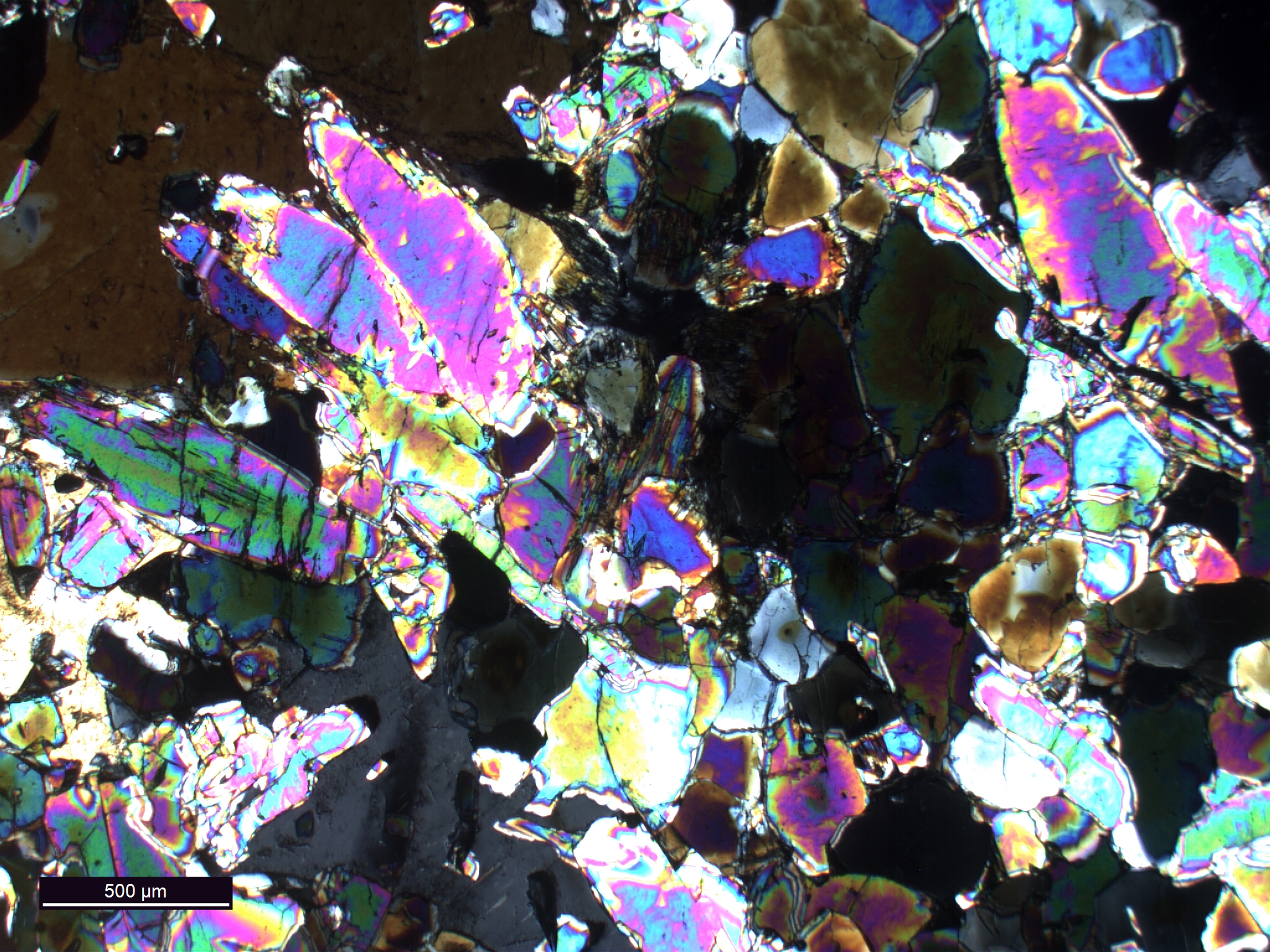
Cordierite displays 1st-order colors up to light yellow. It is biaxial. Rare basal sections may appear pseudohexagonal. It can display polysynthetic twinning (e.g., Fig 6.3.1).
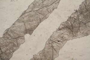
Cordierite is generally anhedral. Cyclic twins can give basal sections a pseudohexagonal appearance. Longitudinal sections may show twin lamellae similar to plagioclase. Irregular, pinching-out, twinning is a diagnostic property for cordierite (e.g., Fig 6.3.6), but sometimes twins do not pinch out and cordierite looks like twinned plagioclase.
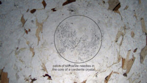
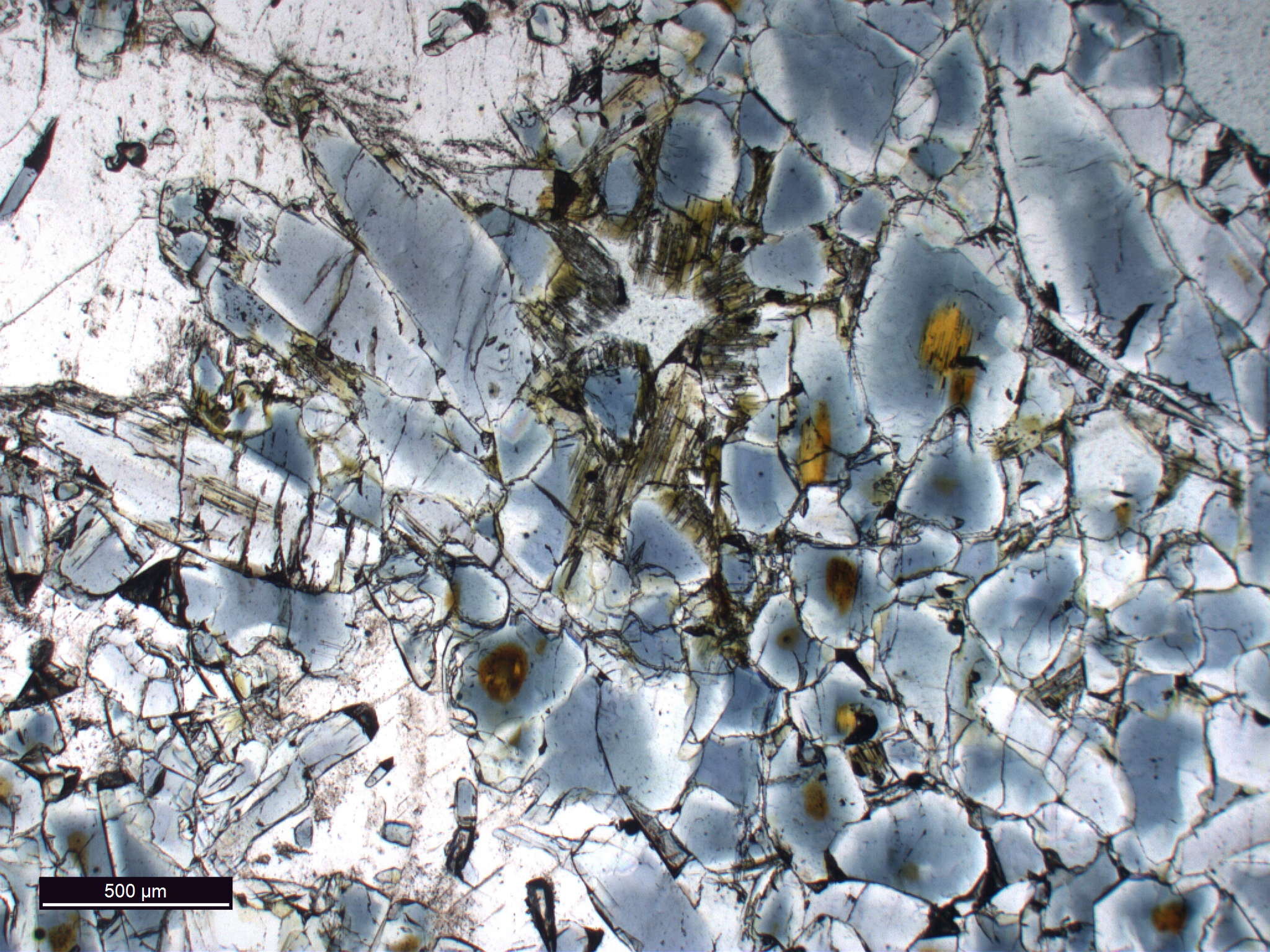
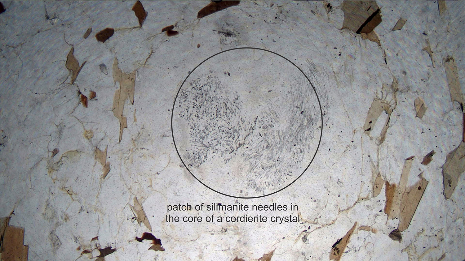
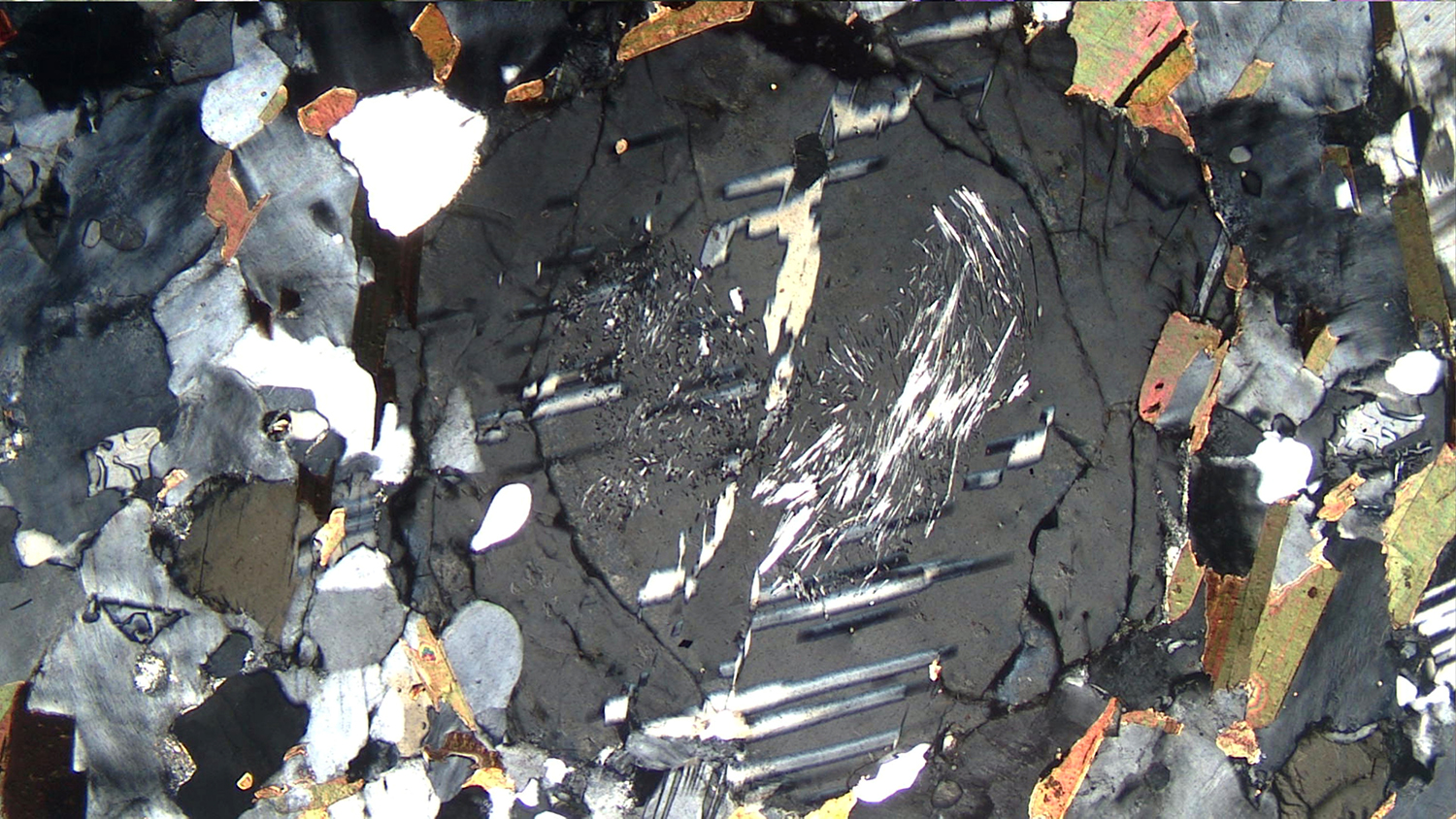
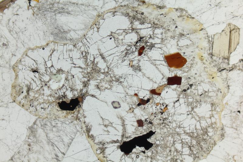
Cordierite’s most distinctive feature is pleochroic halos around zircon and monazite inclusions (Fig 6.3.2). No other mineral shows this. Cordierite has been observed to contain sillimanite needles (Fig 6.3.3) and commonly coexists with sillimanite (and biotite). It commonly alters to pinite, a darkish yellow, gray or green material consisting mostly of fine-grained chlorite, but also muscovite and in some cases clay (Fig 6.3.5). Pinite is typically black in crossed polars and cross-cuts cordierite.
Cordierite has one fair and one poor cleavage, but these are rarely obvious in thin sections.
Similar Minerals—Cordierite is easily mistaken for quartz, but quartz is uniaxial (+) and does not alter. Twinned cordierite may look like plagioclase, but cordierite twins tend to be irregularly spaced and may be discontinuous (Fig 2.10.3). Cordierite can also be spatially associated with other minerals, e.g., forming coronas around garnet or sillimanite.
Optical Properties
■■ Orthorhombic; biaxial (-) or, more rarely (+)
■■ 2V generally 65-90°
■■ α = 1.530-1.560, β = 1.535-1.574, γ= 1.538-1.578; low relief
■■ δ = 0.008-0.018; maximum interference colors are commonly 1st-order white or gray, less commonly yellow
■■ Cordierite is orthorhombic, so exhibits parallel extinction for prismatic grains
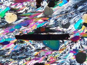
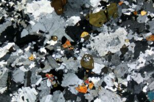
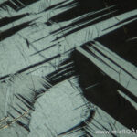
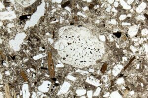
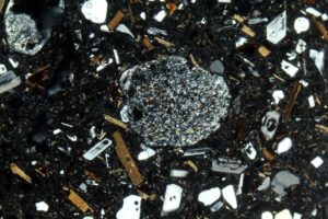
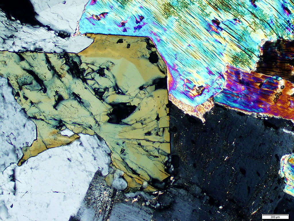
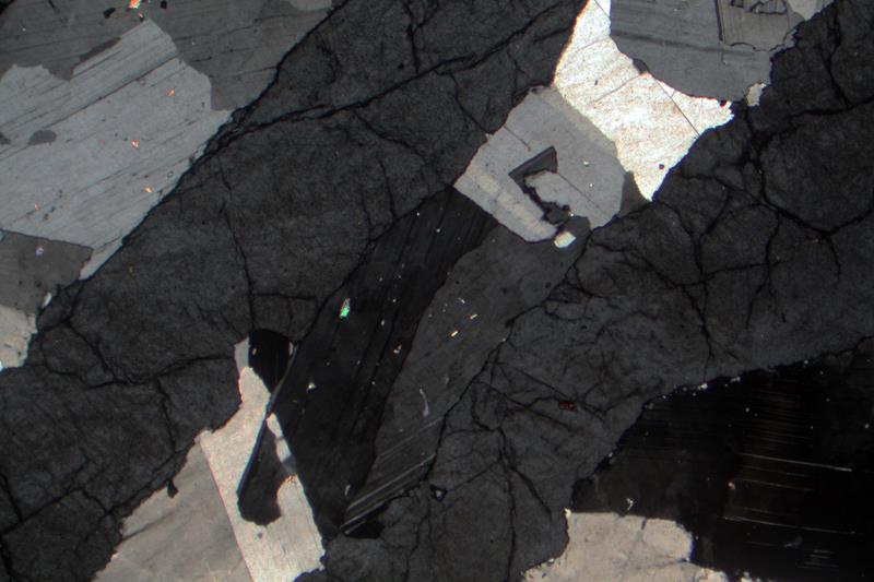
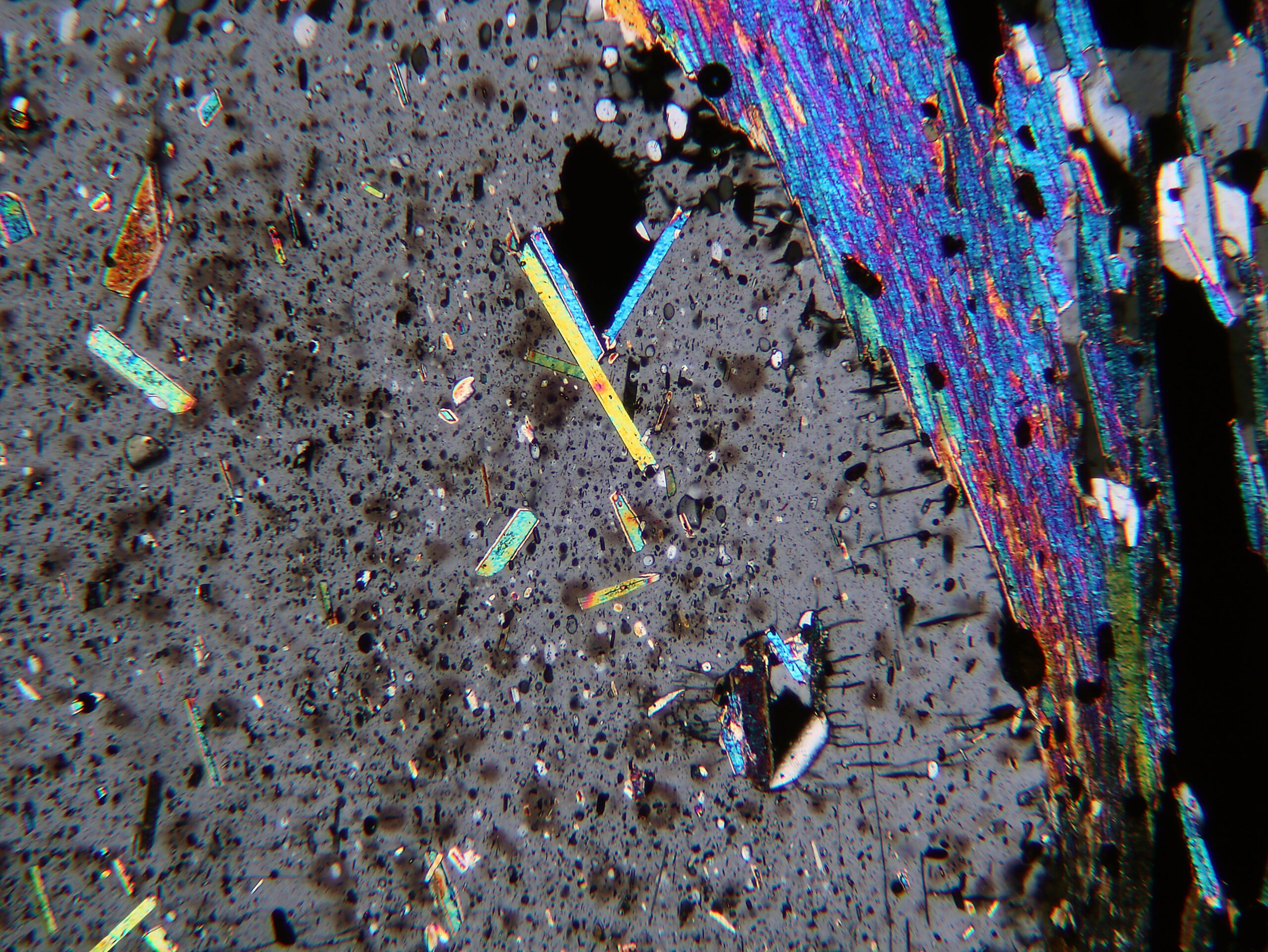
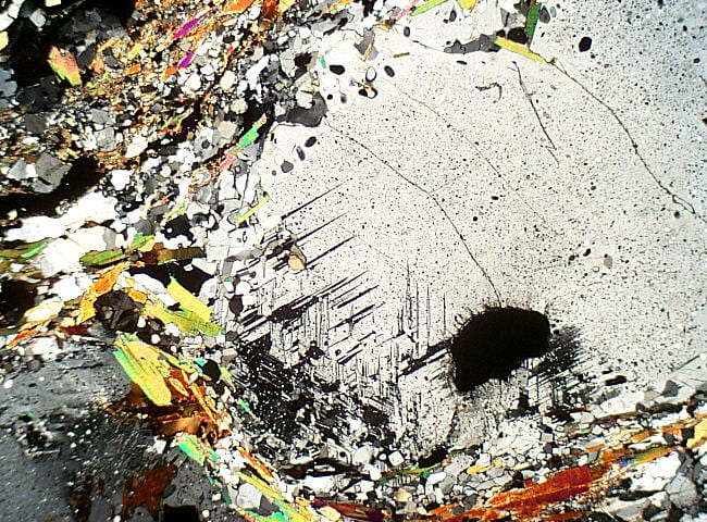
| Fig 6.3.1 Cordierite Gneiss
This spectacular metamorphic rock from near Kazabazua, Quebec, contains abundant low-relief, colorless cordierite and high-relief orthopyroxene, with lesser amounts of moderate-relief, brown to green biotite. The cordierite contains yellow-orange pleochroic halos around radioactive accessory minerals, likely either zircon or monazite. In XP, cordierite has 1st-order gray to white interference colors and is twinned, much like plagioclase. Orthopyroxene also shows 1st-order colors. The halos in cordierite appear as “burn” marks in both PP and XP, and readily distinguish cordierite from plagioclase. FOV = 2.5 mm. |
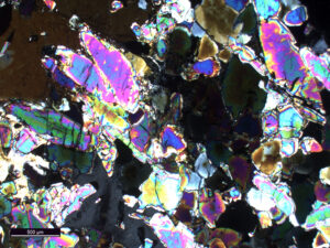
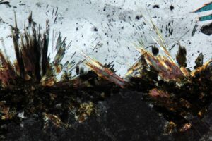
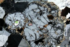
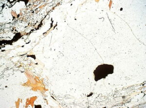
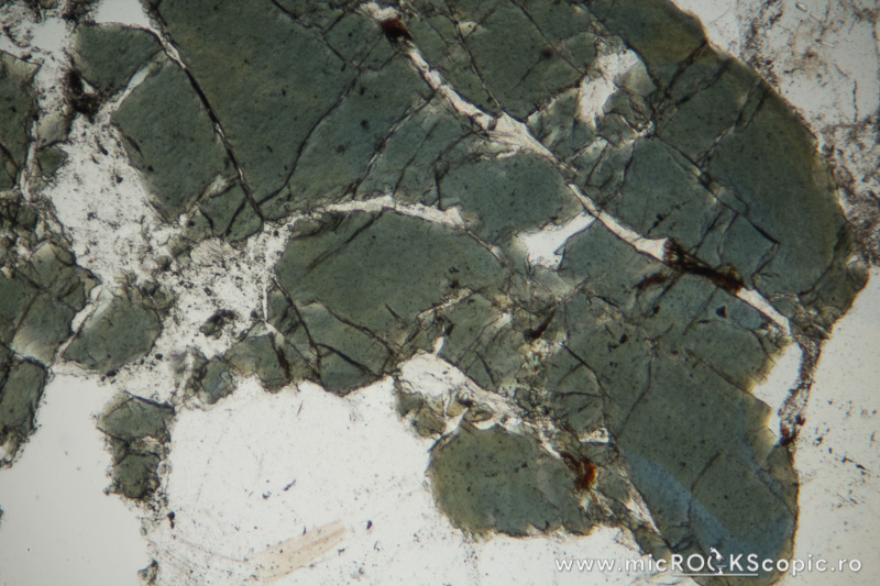
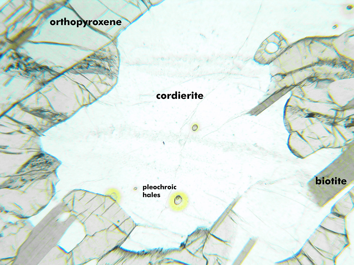
Fig 6.3.2 Cordierite Gneiss
Pleochroic halos, when present, are one of the key diagnostic features for cordierite. The large cordierite grain in the middle of this image contains many yellowish (pleochroic) halos visible in PP around tine inclusions of radioactive minerals, probably monazite or zircon. Colorless elongate flakes of muscovite and quartz are also visible, and opaque grains of various sizes. In XP, the halos, caused by radiation damage, are still weakly visible, colored brown and contrasting with the dark gray 1st-order interference color of the cordierite. The 2nd-order interference colors of muscovite decorate the XP view. This rock comes from southeast Ontario. FOV = 2.5 mm |
|
 |
|
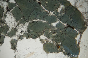
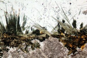
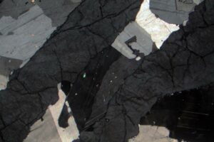
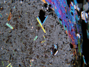
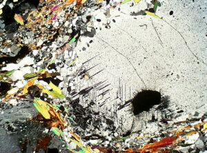
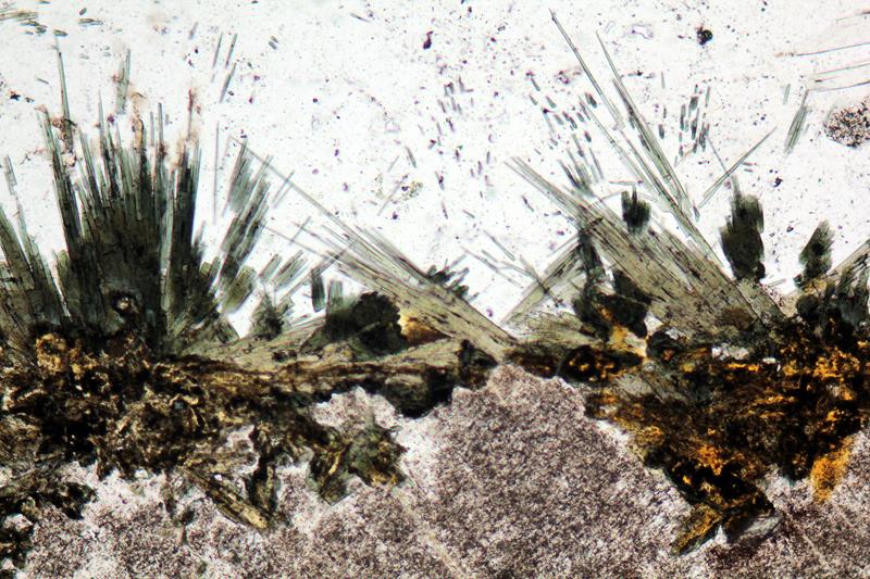
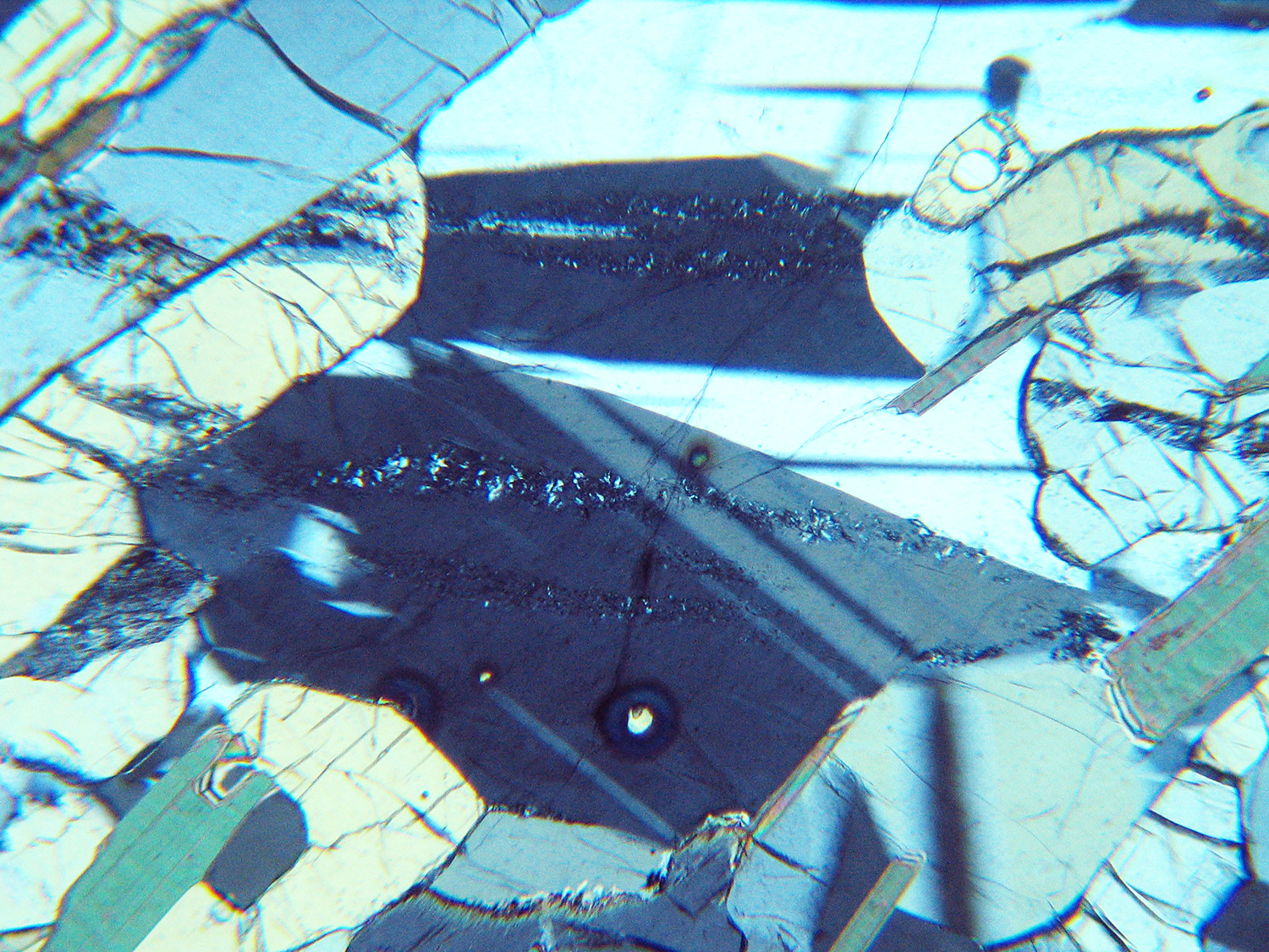
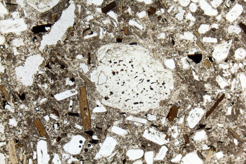
| Fig 6.3.3 Cordierite Gneiss
The center of this view is a single large crystal of cordierite that has a vague hexagonal shape (best visible in the XP view). The crystal contains inclusions of high-relief sillimanite needles (circled in PP). Sillimanite needles commonly occur in cordierite. In crossed polars, the cordierite shows 1st-order gray interference colors and some poorly developed polysynthetic twinning. Other clear grains (PP) are quartz and K-feldspar, but the two are difficult to distinguish. Brown flakes of biotite are also present. The biotite shows up to middle 2nd-order interference colors. This is a cordierite gneiss from Mirond Lake in northern Saskatchewan. FOV = 3.5 mm. |
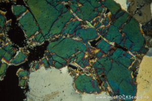
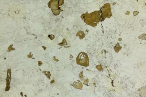
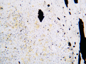
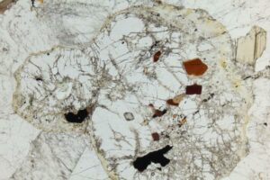
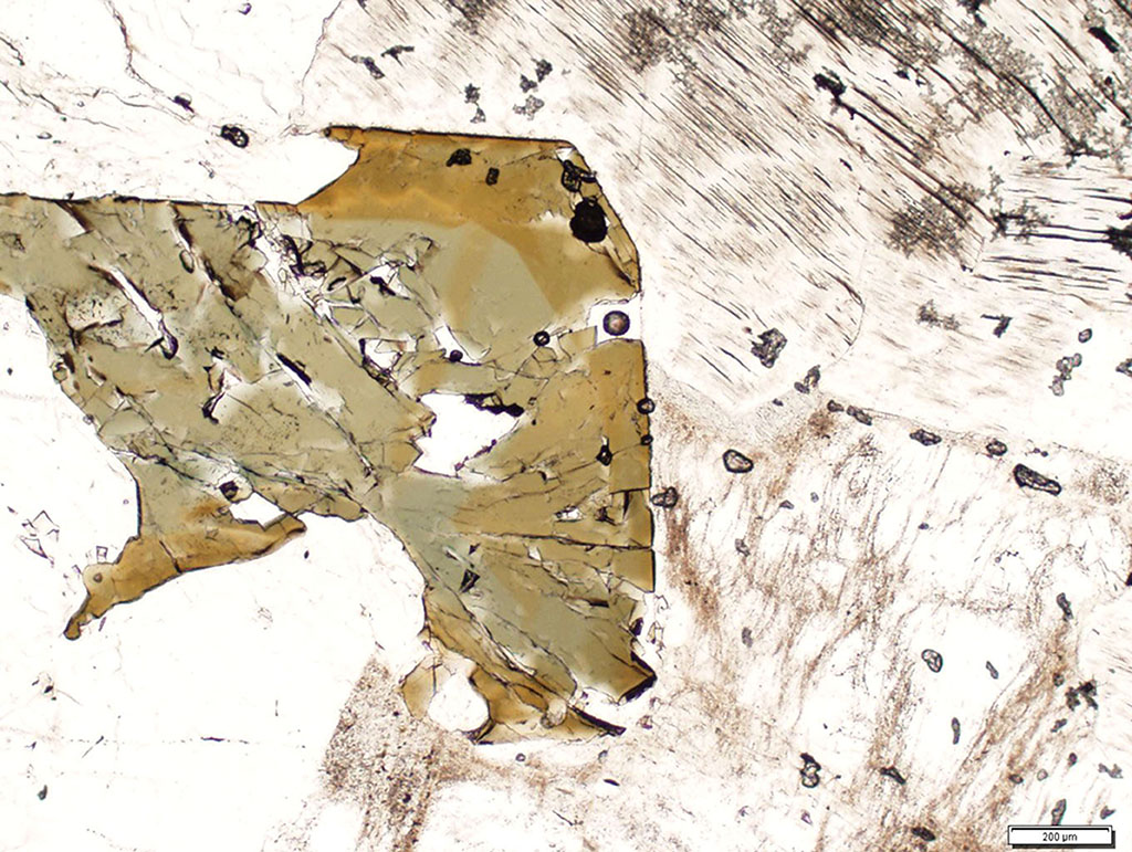
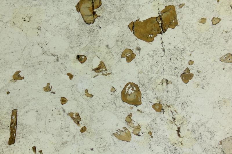
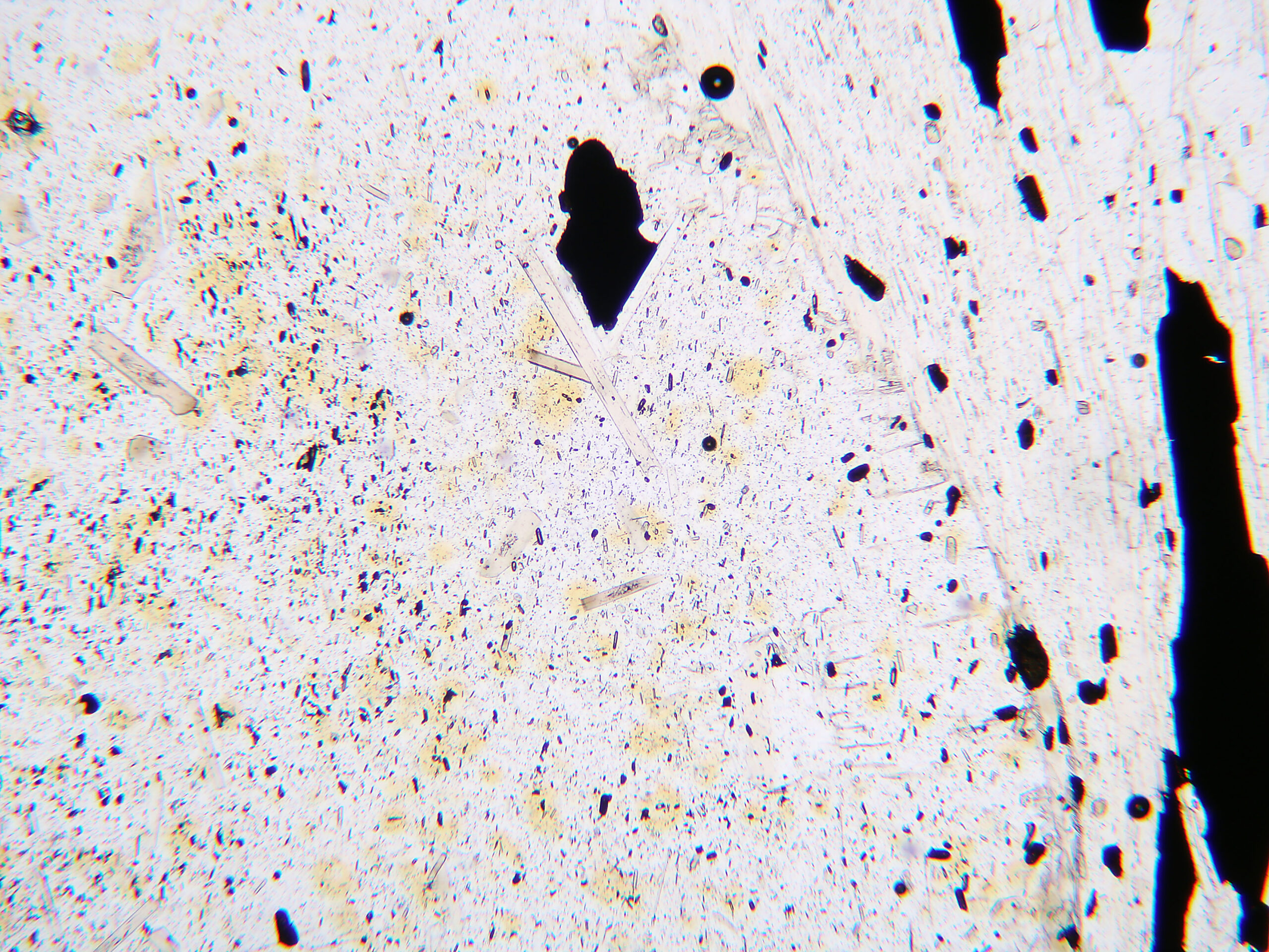
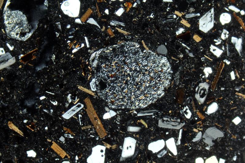
Fig 6.3.4 Cordierite Granite
In this aluminous granite from Elba Island, Italy, a large, low-relief colorless cordierite crystal is moth-eaten and altered along cracks (PP). Clear lower-relief quartz is around the cordierite. Brown biotite and opaque magnetite are also present. In XP, the cordierite crystal is a uniform 1st-order gray. FOV = 7 mm. Photos from www.alexstrekeisen.it. |


